Avicenna Journal of Clinical Microbiology and Infection. 12(4):188-194.
doi: 10.34172/ajcmi.3712
Original Article
Isolation, Molecular Detection, and Sequence Analysis of Pathogenic Leptospira spp. in Paddy Field Water Samples in Amol, Northern Iran
Fereydoun Imani Data curation, Investigation, Methodology, Writing – original draft, 1 
Esmail Fattahi Conceptualization, Methodology, Supervision, Writing – review & editing, 2, *
Rahem Khoshbakht Conceptualization, Methodology, Supervision, Writing – review & editing, 3, 4, *
Sadegh Fattahi Formal analysis, Writing – review & editing, 5
Mohsen Asouri Formal analysis, Writing – review & editing, 6
Author information:
1Department of Microbiology, Am.C., Islamic Azad University, Amol, Iran
2Department of Biology, Am.C., Islamic Azad University, Amol, Iran
3Department of Pathobiology, Amol University of Special Modern Technologies, Amol, Iran
4Zoonotic Diseases Research Group, Faculty of Veterinary Medicine, Amol University of Modern Technologies, Amol, Iran
5North Research Center of Pasteur Institute, Amol, Iran
6Department of Paramedicine, Amol School of Paramedicine, Mazandaran University of Medical Sciences, Sari, Iran
Abstract
Background: Leptospirosis constitutes a zoonotic affliction prevalent in tropical and subtropical locales, instigated by the pathogenic strains of Leptospira. The objective of the present study was to isolate and detect Leptospira from water samples sourced from paddy fields located in Amol, northern Iran.
Methods: A total of 108 samples were procured from rice fields during the spring and summer of 2023. Cultivation, microscopy, and molecular analyses were employed to accurately identify L. interrogans. Both conventional and real-time polymerase chain reactions (RT-PCR) targeting the lipL32 gene were executed for the detection of L. interrogans. In addition, the sequence analysis of the sucA and gluM genes was performed after PCR.
Results: The findings revealed that 6/108 (5.55%) of the samples were culture-positive, with all cases corroborated through both RT-PCR and traditional PCR methods. A simple PCR assay showed positive results for 17/108 (15.74%) water samples. RT-PCR successfully identified 17/108 (15.74%) samples as positive for L. interrogans. The sequence analysis of the sucA gene from the water sample demonstrated high similarity to Leptospira weilii. Eventually, the sequence analysis of the gluM gene from two water samples displayed high similarity to Leptospira borgpetersenii.
Conclusion: This investigation highlights the efficacy of synergizing molecular techniques with traditional culture methodologies for the surveillance of Leptospira in areas characterized by an elevated risk. The current research serves as the inaugural report, delineating the emergence of leptospires from rice field samples, alongside the characterization of isolates obtained from culture.
Keywords: Leptospira, Cultivation, Real-time PCR, Water samples
Copyright and License Information
© 2025 The Author(s); Published by Hamadan University of Medical Sciences.
This is an open-access article distributed under the terms of the Creative Commons Attribution License (
https://creativecommons.org/licenses/by/4.0), which permits unrestricted use, distribution, and reproduction in any medium, provided the original work is properly cited.
Please cite this article as follows: Imani F, Fattahi E, Khoshbakht R, Fattahi S, Asouri M. Isolation, molecular detection, and sequence analysis of pathogenic leptospira spp. In paddy field water samples in Amol, northern Iran. Avicenna J Clin Microbiol Infect. 2025;12(4):188-194. doi:10.34172/ajcmi.3712
Introduction
Leptospirosis is a significant zoonotic disease globally, primarily caused by the pathogenic Leptospira species. It is a severe health risk, particularly in the tropics and subtropics, where environmental and occupational exposure to contamination is high (1). This disease is characterized by a wide spectrum of clinical manifestations, ranging from nonspecific flu-like illness to life-threatening conditions, such as Weil’s disease and pulmonary hemorrhage syndrome (2). The economic burden of the disease, in addition to its association with floods, agriculture, and poor sanitation, underscores its status as a neglected tropical disease (3).
Water is one of the most significant environmental reservoirs of Leptospira, especially in agricultural settings, such as rice paddies, which consist of stagnant water and appropriate climatic conditions for the survival and propagation of these bacteria (4). Rice fields are more relevant in northern Iran, where rice cultivation is one of the most prevalent agricultural activities. Farmers and workers are frequently exposed to water contaminated by infected animal urine, increasing their risk of leptospirosis (5). Monitoring such water sources is critical not only for understanding the local epidemiology of the disease but also for designing effective preventive strategies.
Despite the importance of surveillance, the diagnostic methods for Leptospira have some limitations. The standard method for the clinical identification of leptospirosis is the microscopic agglutination test; however, it is less ideal for environmental samples due to the complexity of water contaminants and the need for live cultures. Traditional culture methods, while required for the isolation of viable organisms, are time-consuming with low sensitivity. Dark-field microscopy is a quicker, though non-specific, alternative (6). Molecular methods, in particular polymerase chain reaction (PCR)-based assays, revolutionized Leptospira detection. Real-time PCR (RT-PCR) targeting the lipL32 gene, a conserved target within pathogenic species, provides sensitive and specific Leptospira interrogans detection directly from environmental samples (7). As Amol is an agriculturally significant region and there is a great possibility of leptospirosis, the current research attempted to isolate and identify L. interrogans in the water samples of paddy fields in the region. Through the combination of culture, molecular, and genotyping methods, the research proposes to provide detailed knowledge on the prevalence, distribution, and genetic diversity of Leptospira in the high-risk area. The findings will not only contribute to what is known about leptospirosis in northern Iran but also to the global effort in the control of this neglected yet significant zoonotic disease.
Methods
Study Area and Sample Collection
Overall, 108 samples were collected during summer and autumn of 2023. In Amol, Mazandaran province, Iran, which is an area well-known for large rice paddies and cattle rearing, sampling was performed from paddy fields with the following sampling conditions:
Water was collected in 108 rice fields in the study area. Approximately 500 mL of surface water was pulled in sterile bottles at multiple locations in each field to collect a representative sample. The sample was cooled at 4°C and sent to the laboratory immediately.
Culture and Microscopy
An approximate volume of 1 mL from each water sample was inoculated into the modified Ellinghausen McCullough Johnson and Harris (EMJH) medium, which was fortified with 5-fluorouracil, rifampicin, and neomycin. The remaining urine and water specimens underwent centrifugation at a force of 3500 g for 15 minutes. Approximately 1 mL of the resultant pellet was subsequently employed for a 10-fold serial dilution within the modified liquid EMJH medium. The inoculated EMJH media were incubated at temperatures ranging from 28 °C to 30 °C over a span of 28 days, with observations for Leptospira-like microorganisms conducted on a weekly basis utilizing a dark field microscope. Positive samples underwent filtration through a 0.2-μM membrane filter, and 100 μL of the filtrate was aliquoted onto EMJH solid medium Leptospira Vanaporn Wuthiekanun agar plates for the purpose of isolation, in accordance with previously established methodologies (8-10). Successfully isolated leptospiral cultures were subsequently transferred into sterile cryovials containing a glycerol solution specifically designed to mitigate freeze damage. These cryovials were then stored in liquid nitrogen tanks at -80°C for extended preservation.
Deoxyribonucleic Acid Extraction
DNA was purified from water employing the QIAamp DNA Mini Kit (QIAGEN, Germany) according to the manufacturer’s instructions with accuracy. Purified DNA was then preserved at −20 °C until subsequently evaluated by PCR. All isolated DNA specimens were adjusted to a standardized concentration of 10–20 ng/μL.
Molecular Detection by Real-Time Polymerase Chain Reaction
To support the amplification of the DNA, an RT-PCR was performed with primers that were specifically created for amplifying the lipL32 gene that is associated with pathogenic Leptospira (1). The assay was further optimized through the incorporation of reference samples (L. interrogans serovar Icterohaemorrhagiae) that had been previously stored at the Pasteur Institute of Iran (North branch). The PCR amplification procedure was executed within a total reaction volume of 25 μL, which comprised 1 × Maxima® SYBR Green I/ROX qPCR master mix (Thermo Scientific, Germany), inclusive of Maxima® Hot Start Taq DNA polymerase, and deoxynucleotide triphosphates within a meticulously optimized PCR buffer that attained a final concentration of 2.5 mM MgCl2. Both forward and reverse primers were utilized to reach the ultimate concentration of 0.4 μM each, succeeded by the addition of 10 μL of extracted DNA (the sample). Each PCR run incorporated negative controls, wherein 10 μL of nuclease-free distilled water was employed in place of the template DNA. L. interrogans serovar Icterohaemorrhagiae DNA ( ≤ 100 ng, 10 μL) served as the positive control. The PCR was performed on the Rotor Gene Q 2plex HRM system (Qiagen, Germany) with an initial denaturation at 95 °C for 10 minutes, followed by 40 cycles of denaturation at 95 °C for 15 seconds, annealing at 53 °C for 30 seconds, extension at 72 °C for 30 seconds, and an ending incubation step at 72 °C for 7 minutes. Melting temperature analysis (70–94°C with readings every 0.5 °C) was performed following a cooling step of 30 °C for 1 minute, according to the manufacturer’s instructions. The cutoff threshold was determined at a threshold cycle of 35, which, based on our empirical observations, represented the final cycle completely devoid of background interference. All experimental protocols were replicated a minimum of two times to ensure the reliability of the results (9,11).
Conventional Polymerase Chain Reaction
A set of primers was designed and utilized to selectively amplify the lipL32 genes within pathogenic Leptospira. Positive control was L. interrogans serovar Icterohaemorrhagiae. The reaction mixtures of 25 µL consisted of 5 µL 5x PCR buffer, 2.0 mM MgCl2, 0.4 µM primer each, 0.2 mM deoxynucleotide triphosphate mix, 1.25 units Taq DNA polymerase (SinaClone, Iran), and 5 µL DNA template. The PCR cycling conditions included primary denaturation at 95 °C for 2 minutes, and then 35 cycles of denaturation at 95 °C for 1 minute, primer annealing at 55 °C for 30 seconds, and extension at 72 °C for 1 minute, followed by a final extension at 72 °C for 5 minutes, and finally ending with an open holding time at 4 °C. Electrophoresis was subsequently run on a 1.5% agarose gel in 1x Tris-acetate-ethylenediaminetetraacetic acid buffer.
Primer Design and Sequence Analysis of the gluM and sucA Genes
Two Leptospira isolates (W7 and W36, samples with good results in RT-PCR and culture) obtained from cultured water samples collected from rice paddies were selected for the molecular analysis of the gluM gene involved in the biosynthesis of extracellular polysaccharides and the sucA gene that encodes the E1 component of the 2-oxoglutarate dehydrogenase complex, which plays a key role in the tricarboxylic acid cycle and bacterial energy metabolism. DNA was extracted using a commercial extraction kit (Yekta Tajhiz, Iran), following the manufacturer’s instructions. The PCR amplification of the gluM and sucA genes was performed using specific primers (Table 1) under standard cycling conditions. Primers for PCR were designed in the present study using Oligo primer analysis software (version 7). The amplified PCR products were visualized by electrophoresis on a 1.5% agarose gel stained with safe stain. Positive PCR amplicons were purified and sent for sequencing (Pishgam Biotech Company, Iran). The recovered nucleotide sequences were compared with known sequences within the NCBI GenBank database using the BLAST algorithm to confirm identity and similarity. Multiple sequences were then aligned using ClustalW, and a phylogenetic tree was constructed from the neighbor-joining method using MEGA software (version X) to determine the evolutionary relationship of the isolates with reference strains (12).
Table 1.
Nucleotide Sequences Used as Primers in PCR and Real-Time PCRs
|
Target gene
|
Sequence (5' to 3')
|
Annealing (°C)
|
Product size (bp)
|
Reference
|
lipL32
(genus/PCR) |
F: CCCAGGGACAAAAACCGTAA
R: ATTTGAGTGGATCAGCGGGC |
55 |
622 |
This study |
lipL32
(genus/real-time PCR) |
F: AAGCATTACCGCTTGTGGTG
R: GAACTCCCATTTCAGCGAT |
53 |
242 |
(11) |
|
gluM (sequencing) |
F: TGTGGTATTAGCCGCAGGGAAG
R: GCAAATACAGCAATGTCTCCGTTC |
56 |
755 |
This study |
|
sucA (sequencing) |
F: TCAGATGGACGGGCTCGTGATC
R: GCAGGCTCGTCCGTTTCATTATG |
58 |
657 |
This study |
Note. PCR: Polymerase chain reaction; F: Forward; R: Reverse.
Results
Results of the Culture and Isolation
Among 108 water samples taken from rice and paddy fields in Amol, 6 (5.55%) were detected as positive by the culture method and showed the presence of the spiral-shaped microorganism in a dark-field microscope image (Figure 1 and 2). All samples that were positive in the culture method were tested using confirmatory PCR, and the presence of the microorganism was confirmed accordingly. Based on the results, no colonies were observed on solid media.

Figure 1.
Dark Field Microscope Image of a Leptospira Isolate Obtained From a Paddy Field Water Sample
.
Dark Field Microscope Image of a Leptospira Isolate Obtained From a Paddy Field Water Sample
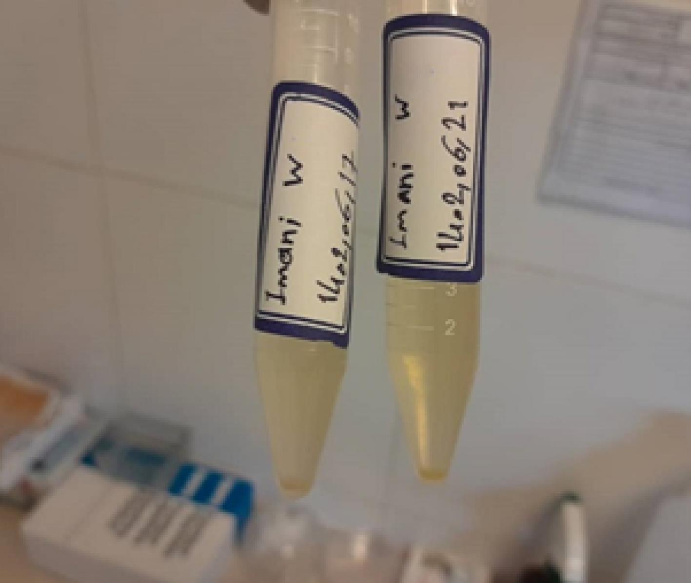
Figure 2.
Growth of Leptospira spp. Isolated From Paddy Field Water Samples in the EMJH Semi-solid Medium. Note. EMJH: Ellinghausen McCullough Johnson and Harris
.
Growth of Leptospira spp. Isolated From Paddy Field Water Samples in the EMJH Semi-solid Medium. Note. EMJH: Ellinghausen McCullough Johnson and Harris
Molecular Detection Results
Among 108 water samples obtained from rice and paddy fields, 17 (15.74%) were detected positive for the presence of pathogenic Leptospira spp. (L. interrogans) by the PCR method targeting the lipL32 gene (Figure 3). In addition, among all water samples taken from rice and paddy fields, 17 (15.74%) were positive for the presence of Leptospira interrogans by the RT-PCR method. The same samples that tested positive in the PCR method also tested positive in the RT-PCR assay. Further, all culture-positive samples demonstrated positive molecular detection in both direct PCR and direct RT-PCR assays.
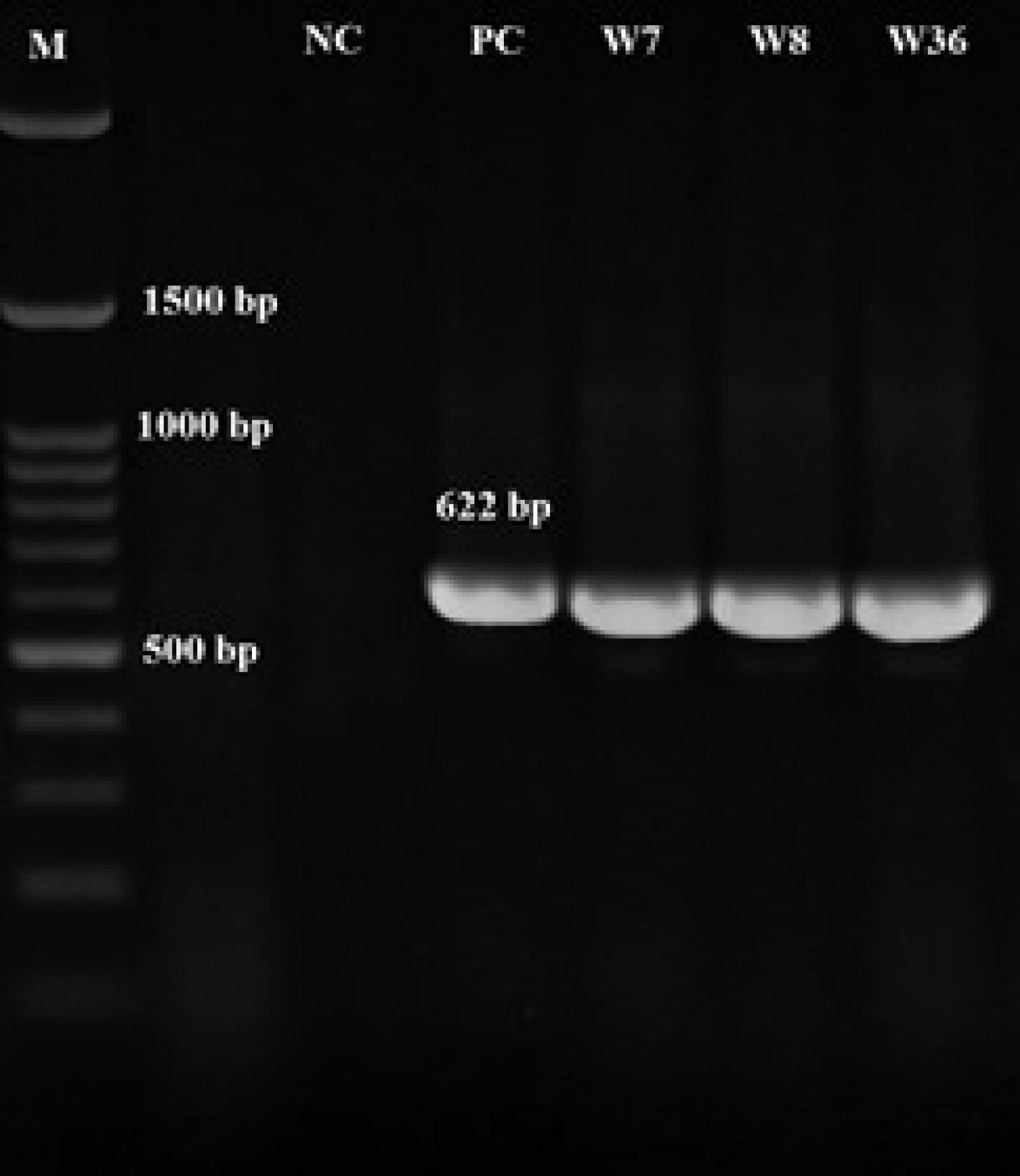
Figure 3.
Agarose Gel Electrophoresis of PCR Products Amplified Using lipL32 gene-Specific Primers From Leptospira spp. Isolated From Paddy Field Water Samples. Note. PCR: Polymerase chain reaction
.
Agarose Gel Electrophoresis of PCR Products Amplified Using lipL32 gene-Specific Primers From Leptospira spp. Isolated From Paddy Field Water Samples. Note. PCR: Polymerase chain reaction
Sequencing Results
Figures 4 and 5 illustrate the electrophoresis results of the PCR product of genes sucA and glmU, respectively. The sequence results of the sucA gene (for one isolate: W7) and the glmU gene (for two isolates: W7 and W36) were registered to the NCBI, and the accession numbers PV613208 (W7-sucA), PV613209 (W36-glmU), and PV613210 (W7-glmU) were received accordingly. Figures 6 and 7 display the phylogenetic tree resulting from the analysis and the comparison of the obtained sequences with the GenBank similar sequences. The high similarity of the sucA gene sequence with Leptospira weilii and the similarity of the gluM gene sequence with Leptospira borgpetersenii were identified.
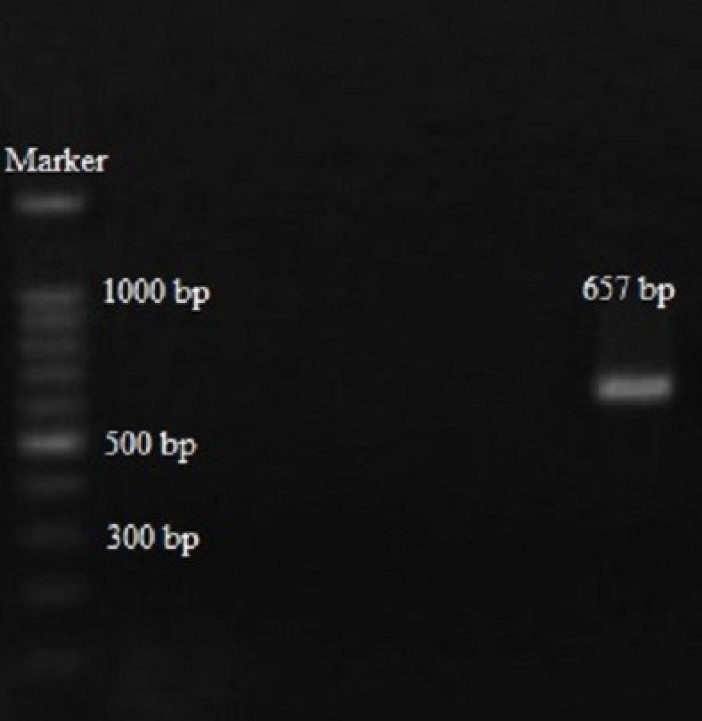
Figure 4.
Agarose Gel Electrophoresis of PCR Products Amplified Using sucA gene-Specific Primers From Leptospira spp. Isolated From Paddy Field Water Samples. Note. PCR: Polymerase chain reaction
.
Agarose Gel Electrophoresis of PCR Products Amplified Using sucA gene-Specific Primers From Leptospira spp. Isolated From Paddy Field Water Samples. Note. PCR: Polymerase chain reaction
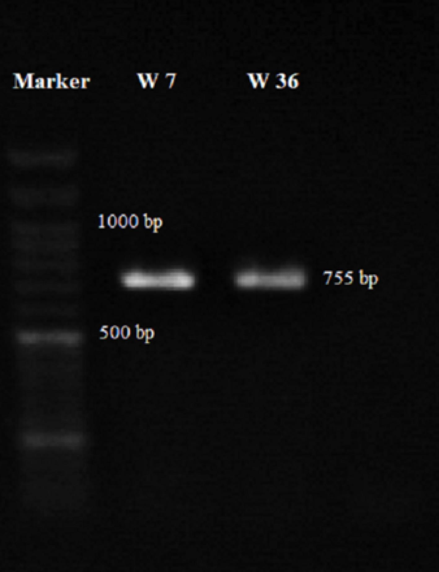
Figure 5.
Agarose Gel Electrophoresis of PCR Products Amplified Using glmU Gene-Specific Primers From Leptospira spp. Isolated from paddy field water samples. Note. PCR: Polymerase chain reaction
.
Agarose Gel Electrophoresis of PCR Products Amplified Using glmU Gene-Specific Primers From Leptospira spp. Isolated from paddy field water samples. Note. PCR: Polymerase chain reaction

Figure 6.
Phylogenetic Dendrogram Generated Using MEGA X Software Based on the sucA Gene Sequences of Leptospira spp. Isolated From Paddy Field Water Samples and Reference Sequences From the NCBI Database. Note. NCBI: The National Center for Biotechnology Information
.
Phylogenetic Dendrogram Generated Using MEGA X Software Based on the sucA Gene Sequences of Leptospira spp. Isolated From Paddy Field Water Samples and Reference Sequences From the NCBI Database. Note. NCBI: The National Center for Biotechnology Information
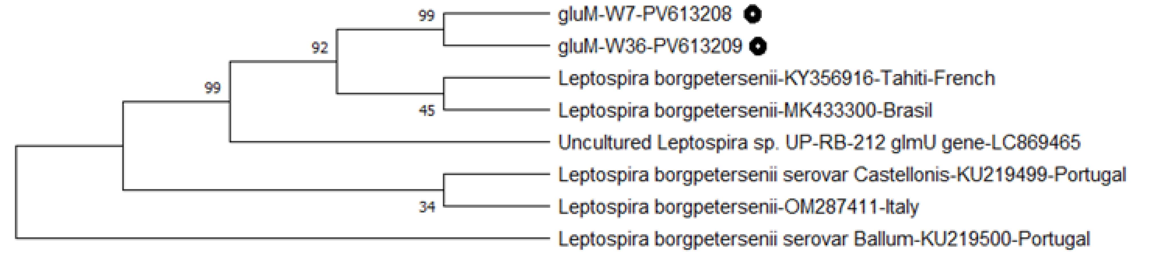
Figure 7.
Phylogenetic Dendrogram Generated Using MEGA X Software Based on the glmU Gene Sequences of Leptospira spp. Isolated From Paddy Field Water Samples and Reference Sequences From the NCBI Database. Note. NCBI: The National Center for Biotechnology Information
.
Phylogenetic Dendrogram Generated Using MEGA X Software Based on the glmU Gene Sequences of Leptospira spp. Isolated From Paddy Field Water Samples and Reference Sequences From the NCBI Database. Note. NCBI: The National Center for Biotechnology Information
Discussion
The present study investigated the prevalence and molecular detection of Leptospira spp. in water samples collected from Amol rice paddies, a region with favorable ecological conditions for the survival of leptospires. Through an integration of traditional culture and molecular techniques, the important prevalence of pathogenic Leptospira species within this habitat was revealed, confirming the public health significance of waterborne leptospirosis in the north of Iran.
While conventional culture is considered the gold standard for Leptospira detection, its sensitivity is limited in environmental samples due to low bacterial load, long incubation time, and high contamination risk (13). In our study, a small subset of water samples (5.55%) yielded positive results through culture, confirmed by dark-field microscopy. These results align with the findings where small numbers of environmental samples were culture-positive despite being collected from high-risk areas (14,15). Ling et al reported that 4.8% of paddy field samples were positive for the presence of Leptospira by both culture and molecular methods (16), which is considerably consistent with the results of the present study. The sampling area in the current study is located in northern Iran and the southern coastal strip of the Caspian Sea, an area that has extensive similarities in terms of climate with Southeast Asia, where many studies show similar prevalence of Leptospira in these areas (17).
However, the isolation of L. interrogans in water samples from paddy fields in Amol highlights the potential public health risk associated with environmental exposure to contaminated surface water, as leptospirosis and rice field fever are commonly present in this region and affect many people every year (5,18). In this region of Iran, paddy fields are located in rural areas that are very close to cities. On the other hand, there are many domestic and wild animals, especially rodents, which are the main carriers of this bacterium and are the main way of transmitting contamination to these waters (19,20).
In this study, by PCR and RT-PCR, 15.74% of the water samples tested positive, indicating the remarkable presence of pathogenic Leptospira species in the rice cultivation areas. These findings conform to those of previous studies conducted in endemic regions, where paddy field environments were identified as major reservoirs for leptospires due to favorable conditions, such as high humidity, standing water, and rodent activity (16,21). The higher detection rate by RT-PCR in comparison to culture (15.74% vs. 5.55%) supports previous findings, indicating that molecular methods are more effective for the direct detection of Leptospira in environmental samples (16). In contrast to cultivation methods, molecular techniques, such as PCR and RT-PCR, offer higher sensitivity and specificity in Leptospira contamination detection and source identification, making them reliable tools for environmental surveillance (22,23). This is particularly important for rapid risk assessment in agricultural communities, where workers are frequently exposed to potentially contaminated water. In addition, many studies emphasize the effectiveness of the molecular identification of Leptospira using the lipL32 gene targeting (24). In the present study, using both PCR and RT-PCR methods, the lipL32 gene was targeted for molecular identification, and acceptable results were obtained accordingly. Given that L. interrogans is one of the most pathogenic species associated with severe human leptospirosis, its detection in agricultural water sources is alarming and warrants targeted interventions, including public health education, rodent control, and possibly vaccination strategies for at-risk populations.
The primary goal of sequencing the Leptospira genes in this study was to perform the multilocus sequence typing technique. Despite the culture and molecular confirmation of the studied samples, the other genes were not well sequenced in the present study. This method was performed in a parallel study on animal isolates and responded well (unpublished data). Therefore, only the sequences that were worth examining were evaluated in this study. Sequence comparison, similar to other methods, confirmed the presence of Leptospira strains, and a significant similarity was observed between L. borgpetersenii and L. weilii in the studied samples. L. borgpetersenii is most commonly associated with domestic animals, such as cattle, and other animals, including rodents (25,26), indicating the contamination of agricultural waters and paddy fields by these animals that roam freely in these areas.
In conclusion, the detection of pathogenic Leptospira in paddy field water through both conventional and molecular approaches highlights the potential public health risk in northern Iran. According to the sequencing results, the presence of genetically diverse species suggests complex transmission dynamics that warrant further investigation. Our findings emphasize the need for environmental monitoring and preventive strategies, particularly for high-risk occupational groups, such as rice farmers.
Conclusion
In conclusion, the detection of pathogenic Leptospira in paddy field water through both conventional and molecular approaches highlights the potential public health risk in northern Iran. According to the sequencing results, the presence of genetically diverse species suggests complex transmission dynamics that warrant further investigation. Our findings emphasize the need for environmental monitoring and preventive strategies, particularly for high-risk occupational groups, such as rice farmers.
Competing Interests
The authors declare that they have no conflict of interests.
Ethical Approval
Essential ethical approval to conduct this study was obtained from the Ethics Committee of Islamic Azad University, Babol Branch, with the code of ethics: IR.IAU.BABOL.REC.1403.133.
Funding
None.
References
- Karpagam KB, Ganesh B. Leptospirosis: a neglected tropical zoonotic infection of public health importance-an updated review. Eur J Clin Microbiol Infect Dis 2020; 39(5):835-46. doi: 10.1007/s10096-019-03797-4 [Crossref] [ Google Scholar]
- Younas K, Saeed L, Umer T, Shah SH, Umar Z, Adil MT, et al. Leptospirosis: a zoonotic disease with reproductive implications. In: Altaf S, Khan A, Abbas RZ, eds. Zoonosis. Vol 4. Faisalabad, Pakistan: Unique Scientific Publishers; 2023. p. 342-55. doi: 10.47278/book.zoon/2023.160.
- Talukder H, Muñoz-Zanzi C, Salgado M, Berg S, Yang A. Identifying the drivers related to animal reservoirs, environment, and socio-demography of human leptospirosis in different community types of southern Chile: an application of machine learning algorithm in one health perspective. Pathogens 2024; 13(8):687. doi: 10.3390/pathogens13080687 [Crossref] [ Google Scholar]
- Antima Antima, Banerjee S. Modeling the dynamics of leptospirosis in India. Sci Rep 2023; 13(1):19791. doi: 10.1038/s41598-023-46326-2 [Crossref] [ Google Scholar]
- Sahneh E, Delpisheh A, Sayehmiri K, Khodabakhshi B, Moafi-Madani M. Investigation of risk factors associated with leptospirosis in the north of Iran (2011-2017). J Res Health Sci 2019; 19(2):e00449. [ Google Scholar]
- Harran E, Hilan C, Djelouadji Z, Ayral F. Epidemiology of leptospirosis: the first literature review of the neglected disease in the Middle East. Trop Med Infect Dis 2022; 7(10):260. doi: 10.3390/tropicalmed7100260 [Crossref] [ Google Scholar]
- Riediger IN, Stoddard RA, Ribeiro GS, Nakatani SM, Moreira SD, Skraba I. Rapid, actionable diagnosis of urban epidemic leptospirosis using a pathogenic Leptospira LipL32-based real-time PCR assay. PLoS Negl Trop Dis 2017; 11(9):e0005940. doi: 10.1371/journal.pntd.0005940 [Crossref] [ Google Scholar]
- Pui CF, Bilung LM, Apun K, Su’ut L. Diversity of Leptospira spp in rats and environment from urban areas of Sarawak, Malaysia. J Trop Med 2017; 2017:3760674. doi: 10.1155/2017/3760674 [Crossref] [ Google Scholar]
- Dorsch R, Ojeda J, Salgado M, Monti G, Collado B, Tomckowiack C. Cats shedding pathogenic Leptospira spp. -An underestimated zoonotic risk? PLoS One 2020; 15(10):e0239991. doi: 10.1371/journal.pone.0239991 [Crossref] [ Google Scholar]
- Wuthiekanun V, Amornchai P, Paris DH, Langla S, Thaipadunpanit J, Chierakul W. Rapid isolation and susceptibility testing of Leptospira spp using a new solid medium, LVW agar. Antimicrob Agents Chemother 2013; 57(1):297-302. doi: 10.1128/aac.01812-12 [Crossref] [ Google Scholar]
- Stoddard RA, Gee JE, Wilkins PP, McCaustland K, Hoffmaster AR. Detection of pathogenic Leptospira spp through TaqMan polymerase chain reaction targeting the LipL32 gene. Diagn Microbiol Infect Dis 2009; 64(3):247-55. doi: 10.1016/j.diagmicrobio.2009.03.014 [Crossref] [ Google Scholar]
- Kumar S, Stecher G, Li M, Knyaz C, Tamura K. MEGA X: molecular evolutionary genetics analysis across computing platforms. Mol Biol Evol 2018; 35(6):1547-9. doi: 10.1093/molbev/msy096 [Crossref] [ Google Scholar]
- Guedes IB, de Souza Rocha K, Negrão MP, de Souza GO, de Paula Castro JF, Cavalini MB. Leptospira transport medium (LTM): a practical tool for leptospires isolation. J Microbiol Methods 2020; 175:105995. doi: 10.1016/j.mimet.2020.105995 [Crossref] [ Google Scholar]
- Nally JE, Ahmed AA, Putz EJ, Palmquist DE, Goris MG. Comparison of real-time PCR, bacteriologic culture and fluorescent antibody test for the detection of Leptospira borgpetersenii in urine of naturally infected cattle. Vet Sci 2020; 7(2):66. doi: 10.3390/vetsci7020066 [Crossref] [ Google Scholar]
- Bierque E, Thibeaux R, Girault D, Soupé-Gilbert ME, Goarant C. A systematic review of Leptospira in water and soil environments. PLoS One 2020; 15(1):e0227055. doi: 10.1371/journal.pone.0227055 [Crossref] [ Google Scholar]
- Ling BL, Tay ZE, Philip N. Detection of Leptospira in environmental samples of wet markets and paddy fields in Penang, Malaysia. Trop Biomed 2025; 42(1):51-7. doi: 10.47665/tb.42.1.009 [Crossref] [ Google Scholar]
- Kesetyaningsih TW, Suryani L, Listyaningrum N. Paddy field area and geographical condition on leptospirosis risk factors in Bantul Regency, Indonesia. E3S Web Conf 2023; 444:02056. doi: 10.1051/e3sconf/202344402056 [Crossref] [ Google Scholar]
- Khalili M, Sakhaee E, Bagheri Amiri F, Asadabadi Safat A, Afshar D, Esmaeili S. Serological evidence of leptospirosis in Iran; a systematic review and meta-analysis. Microb Pathog 2020; 138:103833. doi: 10.1016/j.micpath.2019.103833 [Crossref] [ Google Scholar]
- Boey K, Shiokawa K, Rajeev S. Leptospira infection in rats: a literature review of global prevalence and distribution. PLoS Negl Trop Dis 2019; 13(8):e0007499. doi: 10.1371/journal.pntd.0007499 [Crossref] [ Google Scholar]
- Bradley EA, Lockaby G. Leptospirosis and the environment: a review and future directions. Pathogens 2023; 12(9):1167. doi: 10.3390/pathogens12091167 [Crossref] [ Google Scholar]
- Pui CF, Bilung LM, Su’ut L, Chong YL, Apun K. Detection of Leptospira spp in selected national service training centres and paddy fields of Sarawak, Malaysia using polymerase chain reaction technique. Pertanika J Trop Agric Sci 2017; 40(1):99-109. doi: 10.5555/20173084562 [Crossref] [ Google Scholar]
- Abdul Rahman MS, Khor KH, Khairani-Bejo S, Lau SF, Mazlan M, Roslan MA. TaqMan real-time PCR for detection of pathogenic Leptospira spp in canine clinical samples. J Vet Res 2023; 67(2):187-95. doi: 10.2478/jvetres-2023-0024 [Crossref] [ Google Scholar]
- Loebel P, Azócar-Aedo L, Rodriguez AO, Gallardo MM. Relationship between microscopic agglutination test and real-time PCR assay for detection of seropositivity of pathogenic Leptospira infections in cattle in Chile: a pilot study. Zoonoses 2025; 5(1):983. doi: 10.15212/zoonoses-2025-0003 [Crossref] [ Google Scholar]
- Karunakaran S, Gopinathan GT, Sasidharan H. Taqman based real-time polymerase chain reaction (qPCR) for detection of LipL32 gene of pathogenic leptospires from human, canine, and feline blood samples for diagnosis of acute infections and study carrier status. Pharma Innov J 2022; 11(6):2463-8. [ Google Scholar]
- Moinet M, Wilkinson DA, Aberdein D, Russell JC, Vallée E, Collins-Emerson JM. Of mice, cattle, and men: a review of the eco-epidemiology of Leptospira borgpetersenii serovar Ballum. Trop Med Infect Dis 2021; 6(4):189. doi: 10.3390/tropicalmed6040189 [Crossref] [ Google Scholar]
- Hamond C, Dirsmith KL, LeCount K, Soltero FV, Rivera-Garcia S, Camp P. Leptospira borgpetersenii serovar Hardjo and Leptospira santarosai serogroup Pyrogenes isolated from bovine dairy herds in Puerto Rico. Front Vet Sci 2022; 9:1025282. doi: 10.3389/fvets.2022.1025282 [Crossref] [ Google Scholar]