Avicenna Journal of Clinical Microbiology and Infection. 12(3):102-114.
doi: 10.34172/ajcmi.3662
Original Article
Antibacterial Activity of the Extracts of Dates and Their Leaves Against some Pathogenic Bacterial Isolates
Navid Safa Nova Data curation, Formal analysis, Investigation, Project administration, Validation, Writing – original draft, 1 
Tasnia Ahmed Conceptualization, Formal analysis, Methodology, Resources, Software, Visualization, Writing – original draft, Writing – review & editing, 1, 2, *
Author information:
1Department of Microbiology, Stamford University Bangladesh, Dhaka, Bangladesh
2Faculty of Medicine and Health, School of Clinical Medicine, UNSW Sydney, Kensington, NSW 2052, Australia
Abstract
Background: Many plant-derived natural products, including fruits, fruit skins, seeds, and barks, have been studied for their antibacterial properties. This study was prompted by the global increase in antibiotic-resistant bacterial strains, which pose a growing challenge to global health. Natural phytochemicals are being explored as potential alternatives to conventional antibiotics. This study evaluated the antibacterial activity of five types of dates (Ajwa, Maryam, Sagai, Safawi, and Amber) and date leaves against five clinical isolates, including Escherichia coli, Pseudomonas aeruginosa, Staphylococcus aureus, Klebsiella pneumoniae, and Enterococcus spp.
Methods: To this end, aqueous crude homogenates were prepared by homogenizing fresh samples and tested using the agar well diffusion method. Additionally, sun-dried samples were powdered and extracted using ethanol, methanol, and water. Antibacterial activity was assessed against multidrug-resistant (MDR) clinical isolates.
Results: Among twenty-eight antibiotics tested, P. aeruginosa showed resistance to ten, and Enterococcus spp. to eight. Ethanol and methanol extracts exhibited significantly higher antibacterial activity compared to aqueous crude homogenates and aqueous extracts, with methanol extracts being the most effective. Aqueous extracts demonstrated the least antibacterial potential. Among all tested samples, Amber extracts displayed the highest antibacterial activity, while the other dates represented moderate but comparable results.
Conclusion: The ability of the extracts to inhibit MDR clinical isolates suggests their promising potential as alternative agents for treating infections caused by resistant bacteria.
Keywords: Antibacterial activity, Resistance, Extracts, MIC, MBC
Copyright and License Information
© 2025 The Author(s); Published by Hamadan University of Medical Sciences.
This is an open-access article distributed under the terms of the Creative Commons Attribution License (
https://creativecommons.org/licenses/by/4.0), which permits unrestricted use, distribution, and reproduction in any medium, provided the original work is properly cited.
Please cite this article as follows: Nova NS, Ahmed T. Antibacterial activity of the extracts of dates and their leaves against some pathogenic bacterial isolates. Avicenna J Clin Microbiol Infect. 2025;12(3):102-114. doi:10.34172/ajcmi.3662
Introduction
Date palm (Phoenix dactylifera L.) corresponds to the Arecaceae family, and it is considered a major fruit in the Arabian Peninsula (1,2). Nearly 20 types of dates have been discovered so far, and this fruit is now cultivated in many other countries besides the Arabian Peninsula. Some of these countries include Australia, the United States of America (specifically California and Texas), Mexico, and Southern Africa (1,3). Date palms are considered a nutrition-rich fruit that has also been documented in the Islamic religious book of the Holy Quran (3). It contains many different kinds of vitamins (including vitamins A, C, B1, and B2, and nicotinic acid), minerals (zinc, cadmium, magnesium, sodium, calcium, and potassium), saturated fatty acids (e.g., stearic acid and palmitic acid), unsaturated fatty acids (e.g., linoleic acid and oleic acid), fiber, sugars, and amino acids (4-6). Their high phytochemical content has been linked to various biological properties, including antioxidant, antimicrobial, anti-inflammatory, antiviral, and anticancer effects (7-18).
Treating infectious diseases has become a key alarm in recent years due to the development of multidrug-resistant (MDR) pathogenic microorganisms. Hence, antibiotic therapies often do not work, resulting in high morbidity and mortality rates across the world. Natural therapeutics are being sought to use against such drug-resistant microbes. Plants, fruits, and leaves can contain several distinct phytochemicals that can help combat the resistant forms of microbes (19,20). The antibiotics or any other synthetic drugs take a long time to be completely cleared out from the system after consumption, and occasionally impart some adverse side effects. Natural products are free from such problems, and being inexpensive is an additional benefit (1). The phytochemical content, which is responsible for antimicrobial activity, varies depending on the handling, storage, and extraction method. Thus, the ultimate antimicrobial effects may vary with these differences in processing (21).
Although the antimicrobial potential of date fruits has been reported, studies often focus on a single or a few varieties (e.g., Barhee, Sukri, and Rothana), and very few explore the leaves, which are typically agricultural waste (22). In this study, five commercially important and widely consumed date varieties—Ajwa, Mariam, Amber, Safawi, and Sagai—have been selected along with their leaves. These varieties have been chosen based on their popularity in Middle Eastern traditional medicine, their distinct phytochemical profiles, and limited prior data on their comparative antibacterial efficacy, especially against clinical MDR strains.
This study attempts to uncover the antibacterial activity of five different date palms, namely, Ajwa, Mariam, Amber, Safawi, and Sagai, and the leaves of date palm trees. To our knowledge, this is one of the first comparative studies evaluating both fruit and leaf extracts for their antimicrobial activity against selected clinical MDR bacterial isolates. Extracts prepared using ethanol, methanol, and water were utilized, in addition to the aqueous crude homogenate samples, to determine their antimicrobial potency. The minimal inhibitory concentration (MIC) and minimal bactericidal concentration (MBC) values are assessed following the verification of the antibacterial activity of the extracts against selected MDR bacterial strains obtained from clinical specimens.
Materials and Methods
Study Location and Sampling Procedures
Five pathogenic bacterial isolates (Escherichia coli, Klebsiella pneumoniae, Staphylococcus aureus, Pseudomonas aeruginosa, and Enterococcus spp.) were chosen to determine their inhibition by the antibacterial activity of natural phytochemicals. The antibacterial activity assays were performed using clinical bacterial isolates obtained from our laboratory collection. Standard reference strains (e.g., ATCC strains) were not used due to their unavailability. Five different date samples (Amber, Safawi, Mariyam, Sagai, and Ajwa) and the leaves of the date tree were obtained from various markets across Dhaka, Bangladesh. The research was conducted in the microbiology laboratory at the Department of Microbiology, Stamford University, Bangladesh, between September and December 2020.
Antibiotic Susceptibility Testing of Pathogenic Isolates
A total of twenty-eight frequently prescribed antibiotics were selected to evaluate the sensitivity of the clinical isolates, including amoxicillin (25 µg), azithromycin (15 µg), meropenem (10 µg), ceftazidime (30 µg), ciprofloxacin (5 µg), gentamicin (10 µg), amikacin (30 µg), cefixime (30 µg), cefuroxime (30 µg), cephradine (30 µg), and nitrofurantoin (300 µg). The remaining antibiotics were vancomycin (30 µg), teicoplanin (30 µg), clotrimazole (30 µg), piperacillin/tazobactam (30 µg), colistin (30 µg), doxycycline (30 µg), fusidic acid (10 µg), amoxiclav (30 µg), imipenem (10 µg), linezolid (30 µg), doripenem (10 µg), tigecycline (15 µg), clindamycin (10 µg), levofloxacin (5 µg), cefepime (30 µg), nalidixic acid (30 µg), and ceftriaxone (30 µg). Antibiotic susceptibility testing was performed using the Kirby-Bauer disc diffusion method (23). The zones of inhibition were measured, and bacterial isolates were categorized as sensitive or resistant according to the Clinical and Laboratory Standards Institute guidelines (24).
Processing of Samples
Upon arrival at the laboratory, the samples were thoroughly rinsed with tap water, followed by multiple washes with distilled water to remove salts, soil, and other contaminants. Equal weights (10 g) of raw fruits were used for all extractions to ensure standardization. For aqueous crude homogenate preparation, each 10 g sample was homogenized with 90 mL of 0.85% saline solution.
For solvent extraction, the samples were chopped and air-dried at room temperature for 10 days until fully dehydrated. The dried samples were ground into fine powder and stored in airtight containers at room temperature until use. Equal amounts (5 g) of the powdered sample were extracted using different solvents (ethanol, methanol, and distilled water), each at a volume of 100 mL, under identical maceration conditions (72 hours at room temperature with occasional shaking). The extracts were filtered, concentrated under reduced pressure, and stored at 4 °C. The use of a fixed weight-to-volume ratio of 5 g of the powdered sample per 100 mL of the solvent follows commonly accepted protocols for crude plant extract preparation in phytochemical and antimicrobial studies (25). This method ensures consistent extraction conditions across all varieties and solvents. While concentration-based methods are standard for purified compounds, weight-to-volume ratios are preferred for comparative crude extract studies.
Although this study did not include phytochemical screening or standardization, the aim was to conduct a preliminary comparison of the antibacterial activity of the aqueous crude homogenates. Future studies will incorporate phytochemical profiling to help identify bioactive constituents responsible for observed effects.
Antibacterial Activity of Extracts (Crude, Ethanolic, Methanolic, and Aqueous)
Antibacterial activity was assessed using the agar well diffusion method. Positive control (Gentamicin 10 µg/disc) was included for comparison with the extracts, and an appropriate solvent blank (saline) was used as a negative control. The zones of inhibition were measured in mm to compare the efficacy of the extracts with standard antibiotics.
Bacterial suspensions were prepared by inoculating the isolates into normal saline and incubating them at 37 °C until the turbidity matched the McFarland standard (approximately 108 CFU/mL) (26). A bacterial lawn was then created on Mueller-Hinton agar plates using sterile cotton swabs for each bacterial strain separately. The wells were made in the agar, and 100 µL of crude, ethanolic, methanolic, and aqueous date extracts were introduced into the respective wells. The plates were incubated at 37 °C for 24 hours. After incubation, the zones of inhibition around the wells were measured in mm to evaluate antibacterial activity.
Sample sizes varied for each bacterial species, which are detailed in the figure legends (n = 2–5). For instance, K. pneumoniae was represented by two isolates (n = 2), which limits the statistical power for this species.
Positive and negative controls were included only in the agar well diffusion assays to confirm the responsiveness of bacterial isolates and validate the assay conditions. These controls ensured that observed zones of inhibition were attributable to antibacterial activity.
For MIC and MBC assays, controls were not included because these tests aimed specifically to quantify the potency of the crude plant extracts under investigation. Considering that the antibacterial activity of the extracts was already established and validated through the diffusion method with controls, the MIC and MBC determinations focused on measuring extract effectiveness without additional control antibiotics.
Determination of Minimal Inhibitory Concentration and Minimal Bactericidal Concentration
Sample extracts were diluted to concentrations of 500 mg/mL, 250 mg/mL, and 125 mg/mL using sterile nutrient broth. Overall, 0.2 mL of bacterial suspension was added to each dilution tube. The tubes were incubated at 37 °C for 24 hours, and the lowest concentration showing no visible bacterial growth was recorded as the MIC. Subsequently, loopful samples from the clear tubes were streaked onto fresh nutrient agar plates to determine the MBC, identified as the lowest extract concentration where no bacterial growth occurred.
All experiments were performed with biological replicates (n = 2–5, specified in figure legends). The data are presented as means ± standard deviations (SD). Statistical analyses were conducted using RStudio.
To compare inhibition zones among different extracts and bacterial species, one-way analysis of variance (ANOVA) was performed, followed by Tukey’s post-hoc test to identify significant pairwise differences. Where only two groups were compared, differences between extract types and bacterial strains were assessed for MIC and MBC values using ANOVA with appropriate post-hoc tests.
A P value of less than 0.05 was considered statistically significant. All statistical tests were two-tailed.
Results
Antibiotic Resistance Profile
Enterococcus spp. showed resistance to eight antibiotics, while K. pneumoniae exhibited intermediate susceptibility to three and resistance to one. S. aureus was resistant to three antibiotics. P. aeruginosa demonstrated the highest resistance, being resistant to 10 out of the 28 tested antibiotics. E. coli represented resistance to three antibiotics and intermediate resistance to one. Overall, the isolates were MDR, with P. aeruginosa and Enterococcus spp., representing the broadest resistance spectra, while the remaining species retained susceptibility to several antibiotics (Figure 1).
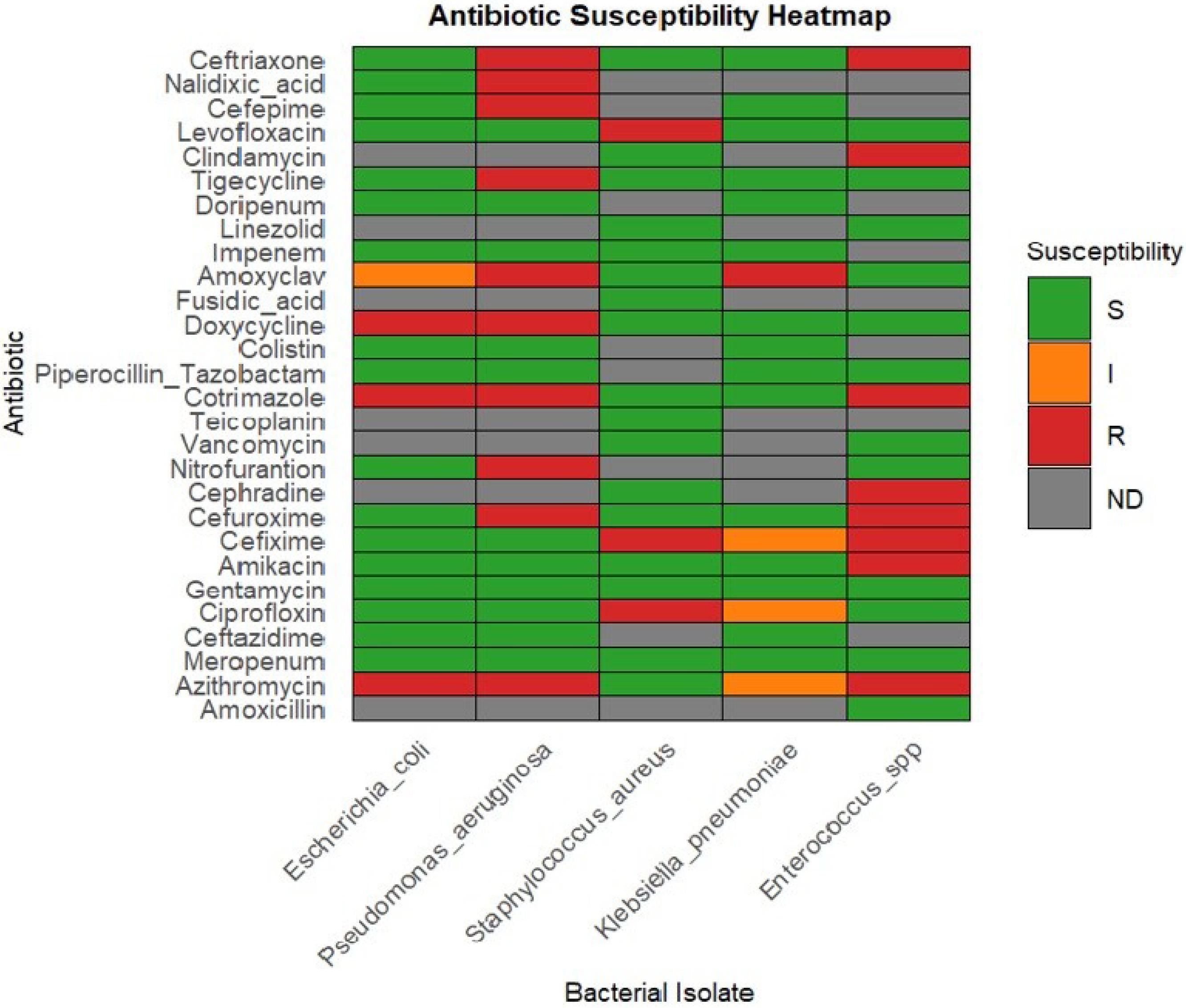
Figure 1.
Antibiotic Susceptibility of Bacterial Isolates
.
Antibiotic Susceptibility of Bacterial Isolates
Antimicrobial Activity – Aqueous Crude Homogenates
The antibacterial activity of aqueous crude homogenates was assessed by measuring the mean zone of inhibition (mm ± SD). For E. coli, Maryam and leaf extracts produced inhibition zones of 12.3 ± 2.08 mm and 12.0 ± 2.00 mm, respectively, compared to 10.3 ± 2.08 mm for Amber; these differences were not statistically significant (P = 0.486). Similar non-significant differences (P = 0.502) were observed for S. aureus across Amber (11.0 ± 1.41 mm), Maryam (10.8 ± 0.96 mm), Sagai (9.5 ± 1.91 mm), and leaf extracts (10.8 ± 1.50 mm). For P. aeruginosa, K. pneumoniae, and Enterococcus spp., only the leaf extract was tested, resulting in moderate inhibition; however, a statistical comparison was limited by small replicate numbers. Overall, no statistically significant differences were found among aqueous crude homogenates (Figure 2).
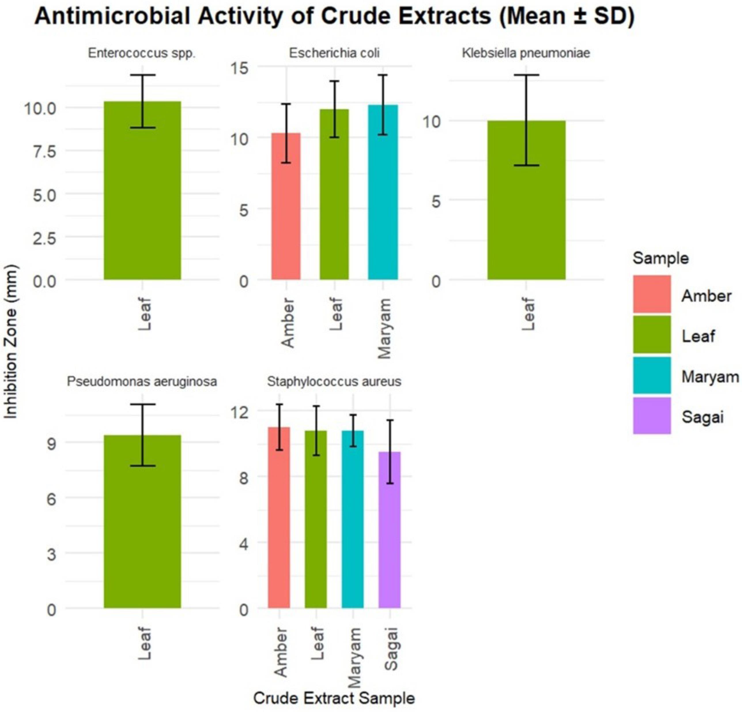
Figure 2.
Antibacterial Activity of Crude Extracts
.
Antibacterial Activity of Crude Extracts
Solvent-Based Extract Efficacy
Ethanol, methanol, and aqueous extracts were further evaluated for antibacterial potency. The ethanol extract of Amber inhibited E. coli with a mean zone of 26.33 ± 3.05 mm, which was the largest inhibition zone observed among ethanol extracts for this species. P. aeruginosa was most inhibited by the methanol extract of Safawi, producing a zone of 12.0 ± 2.83 mm. Aqueous extracts consistently showed low or no inhibition zones ( < 10 mm). For S. aureus, the ethanol extract of Sagai displayed the greatest inhibition zone (13.75 ± 1.26 mm). K. pneumoniae was most susceptible to the Safawi methanol extract, with an inhibition zone of 28.0 ± 2.83 mm. Enterococcus spp. exhibited inhibition zones ranging from 29.0 ± 1.41 mm to 33.0 ± 4.24 mm for ethanol and methanol extracts. No inhibition was recorded for negative controls, validating experimental controls. In general, ethanol extracts, particularly Amber and Sagai, were most effective against E. coli and S. aureus, while methanol extracts—especially Safawi—were effective against P. aeruginosa and K. pneumoniae (Table 1, Figure 3). Biological replicates ranged from 2 to 5, depending on species, with K. pneumoniae represented by only two isolates (n = 2), limiting statistical power for this species.
Table 1.
Antibacterial Effects of Ethanol, Methanol, and Aqueous Extracts (100 µL) From Date Fruit and Leaf Samples Against Pathogenic Bacteria (Zone of Inhibition in mm, Mean ± SD)
|
Samples/Isolates
|
Amber
|
Safawi
|
Maryam
|
Sagai
|
Ajwa
|
Leaf
|
Positive Control
|
Negative Control
|
|
Ethanol
|
Methanol
|
Aquaous
|
Ethanol
|
Methanol
|
Aquaous
|
Ethanol
|
Methanol
|
Aquaous
|
Ethanol
|
Methanol
|
Aquaous
|
Ethanol
|
Methanol
|
Aquaous
|
Ethanol
|
Methanol
|
Aquaous
|
Escherichia coli
(n = 3) |
26.33 ± 3.05 |
10.00 ± 2.00 |
7.67 ± 0.58 |
9.67 ± 2.08 |
10.00 ± 1.00 |
7.67 ± 1.53 |
11.33 ± 2.08 |
9.67 ± 1.53 |
0 |
12.00 ± 2.00 |
9.00 ± 1.00 |
7.33 ± 1.53 |
9.33 ± 1.53 |
13.00 ± 2.65 |
8.33 ± 0.58 |
9.33 ± 2.52 |
11.00 ± 3.00 |
6.33 ± 1.53 |
21.00 ± 3.61 |
0 |
Pseudomonas aeruginosa
(n = 5) |
10.40 ± 1.97 |
10.60 ± 2.45 |
3.80 ± 2.17 |
9.00 ± 2.94 |
3.6 ± 3.51 |
0 |
10.60 ± 2.07 |
12.0 ± 2.83 |
6.67 ± 1.53 |
9 ± 1 |
10 ± 2.16 |
7 ± 1.41 |
11.4 ± 2.19 |
5.4 ± 4.98 |
2.8 ± 3.9 |
7.8 ± 4.49 |
2.6 ± 3.72 |
0 |
20.8 ± 2.28 |
0 |
Staphylococcus aureus
(n = 4) |
12 ± 2.94 |
11.25 ± 1.5 |
3.25 ± 3.77 |
10 ± 1.63 |
9.75 ± 2.06 |
3 ± 3.56 |
11.75 ± 2.75 |
11.25 ± 1.89 |
8 ± 1.63 |
13.75 ± 1.26 |
10.25 ± 2.87 |
9.25 ± 0.95 |
9.75 ± 2.36 |
9.75 ± 2.06 |
7.5 ± 1.29 |
10.75 ± 0.95 |
11.25 ± 2.22 |
3.5 ± 4.12 |
24.25 ± 3.30 |
0 |
|
Klebsiella pneumoniae (n = 2) |
12 ± 2.83 |
28 ± 2.83 |
8.5 ± 2.12 |
13 ± 1.41 |
12.5 ± 3.54 |
7.5 ± 2.12 |
13 ± 1.41 |
0 |
0 |
13 ± 2.83 |
0 |
0 |
14 ± 1.41 |
16 ± 1.41 |
8.5 ± 2.12 |
9 ± 1.41 |
28.5 ± 2.12 |
9.5 ± 2.12 |
29 ± 4.24 |
0 |
|
Enterococcus spp. (n = 2) |
10.5 ± 2.12 |
29 ± 1.41 |
7 ± 1.41 |
13.5 ± 2.12 |
32.5 ± 3.54 |
11 ± 1.41 |
9 ± 1.41 |
13.5 ± 2.12 |
8.5 ± 2.12 |
13.5 ± 2.12 |
29 ± 1.41 |
11.5 ± 2.12 |
16 ± 1.41 |
16.5 ± 2.12 |
9.5 ± 2.12 |
12 ± 2.83 |
9.5 ± 0.71 |
5.5 ± 2.12 |
33 ± 4.24 |
0 |
Note. ***Ethanol-ethanol solvent-based extract, methanol-methanol solvent-based extract, aqueous-water-based extract.
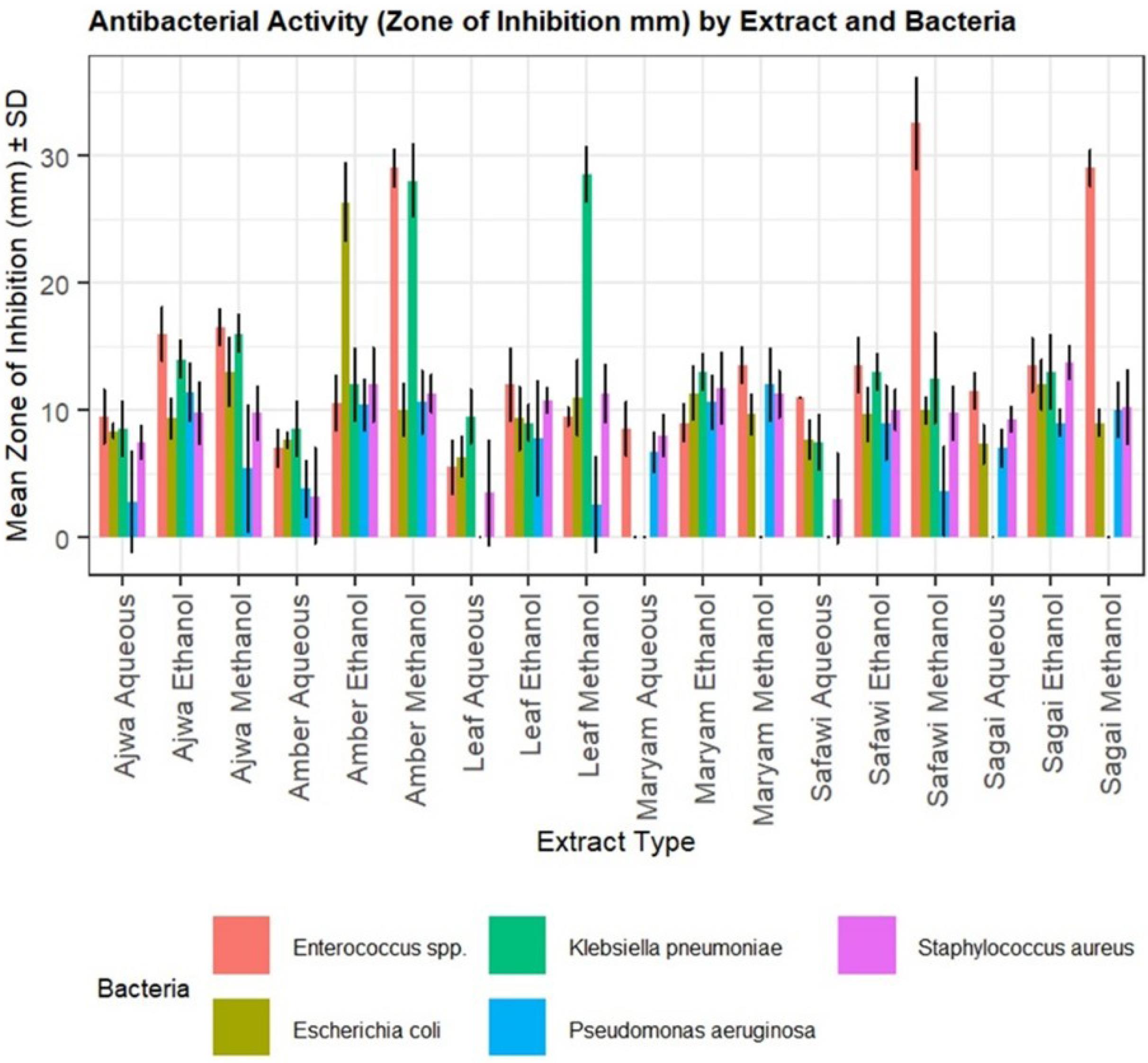
Figure 3.
Antibacterial Activity of Different Extracts Against Different Bacteria
.
Antibacterial Activity of Different Extracts Against Different Bacteria
Antibacterial Activity of Various Date Extracts Against Five Bacterial Species
Bar plots represent the mean zone of inhibition (mm) ± SD for each extract type. The extracts from six types of date samples (Ajwa, Amber, Safawi, Maryam, Sagai, and leaf) were tested using three solvents (ethanol, methanol, and aqueous). The antibacterial effect was assessed against E. coli, P. aeruginosa, S. aureus, K. pneumoniae, and Enterococcus spp. Error bars indicate the SD from biological replicates (n = 2–5). Each bacterial species is color-coded, and legends are arranged below the plot for clarity.
Minimum Inhibitory Concentration
Extract concentrations were evaluated to determine the MIC, defined as the lowest concentration at which no visible turbidity was observed (OD600 ≤ 0.1 or no turbidity visible by eye). For ethanol extracts, the visible inhibition of P. aeruginosa and K. pneumoniae occurred at 500 µg/mL for Amber, and similarly for Ajwa (P. aeruginosa, S. aureus, and Enterococcus spp.). Leaf extracts also inhibited S. aureus and Enterococcus spp. at this concentration. The Sagai and Maryam ethanol extracts inhibited bacterial growth as early as 250 µg/mL (Figure 4). Methanol extracts showed greater potency, with MICs as low as 125 µg/mL for Maryam, Safawi, and Ajwa against E. coli. The same MIC was noted for Sagai (K. pneumoniae, P. aeruginosa, and S. aureus), Maryam (P. aeruginosa), and Ajwa (K. pneumoniae). Notably, P. aeruginosa required a higher MIC (1000 µg/mL) for the Safawi methanol extract (Figure 5). Aqueous extracts generally displayed higher MICs ( ≥ 375 µg/mL) or no inhibition at concentrations up to 1000 µg/mL, particularly for P. aeruginosa with the Safawi extract (Figure 6). Sample sizes for MIC assays are specified in figure legends; K. pneumoniae data were limited (n = 2), which may affect statistical interpretation.
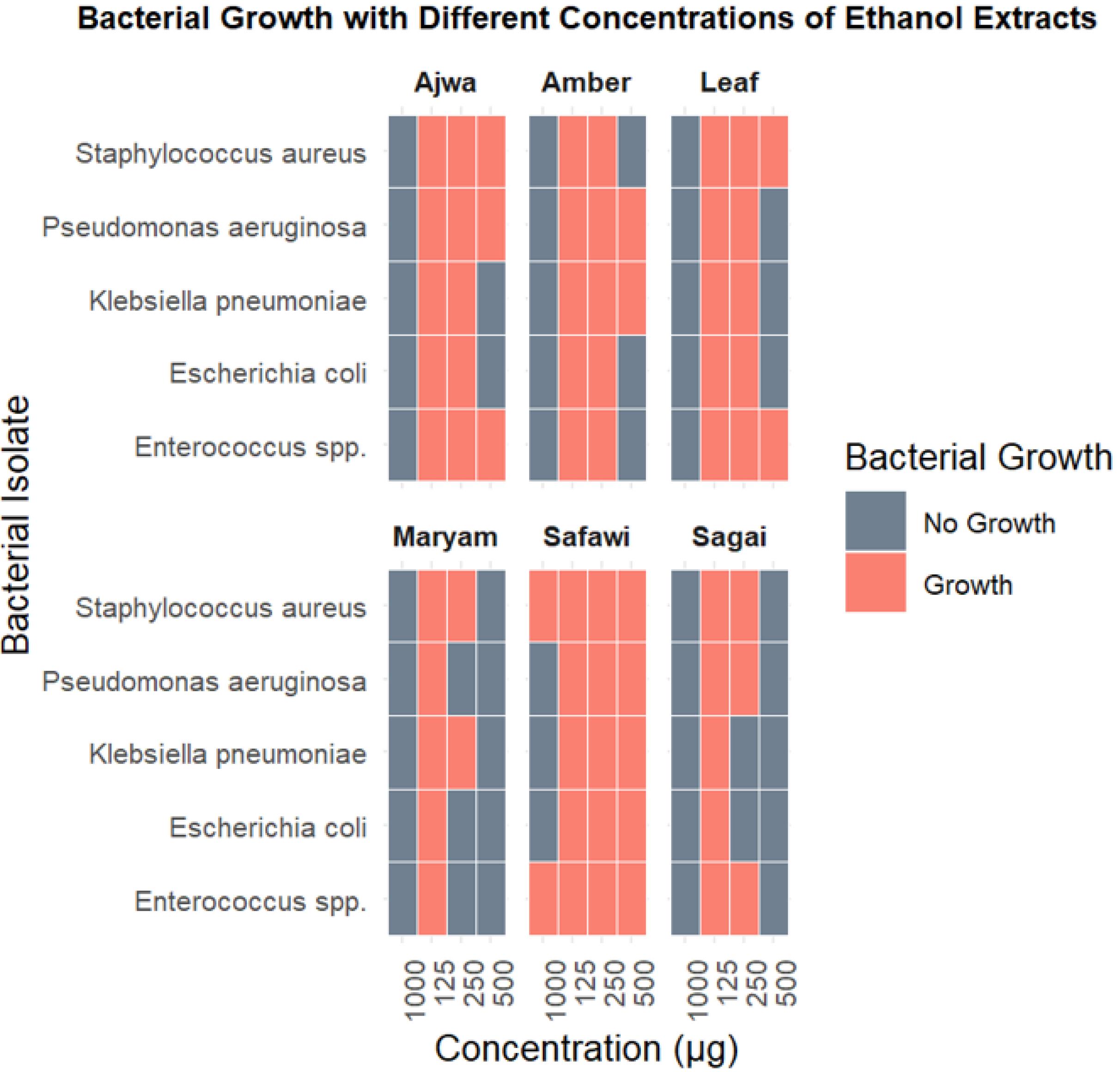
Figure 4.
Bacterial Growth with Different Concentrations of Ethanol Extracts of Date Fruits and Leaves
.
Bacterial Growth with Different Concentrations of Ethanol Extracts of Date Fruits and Leaves
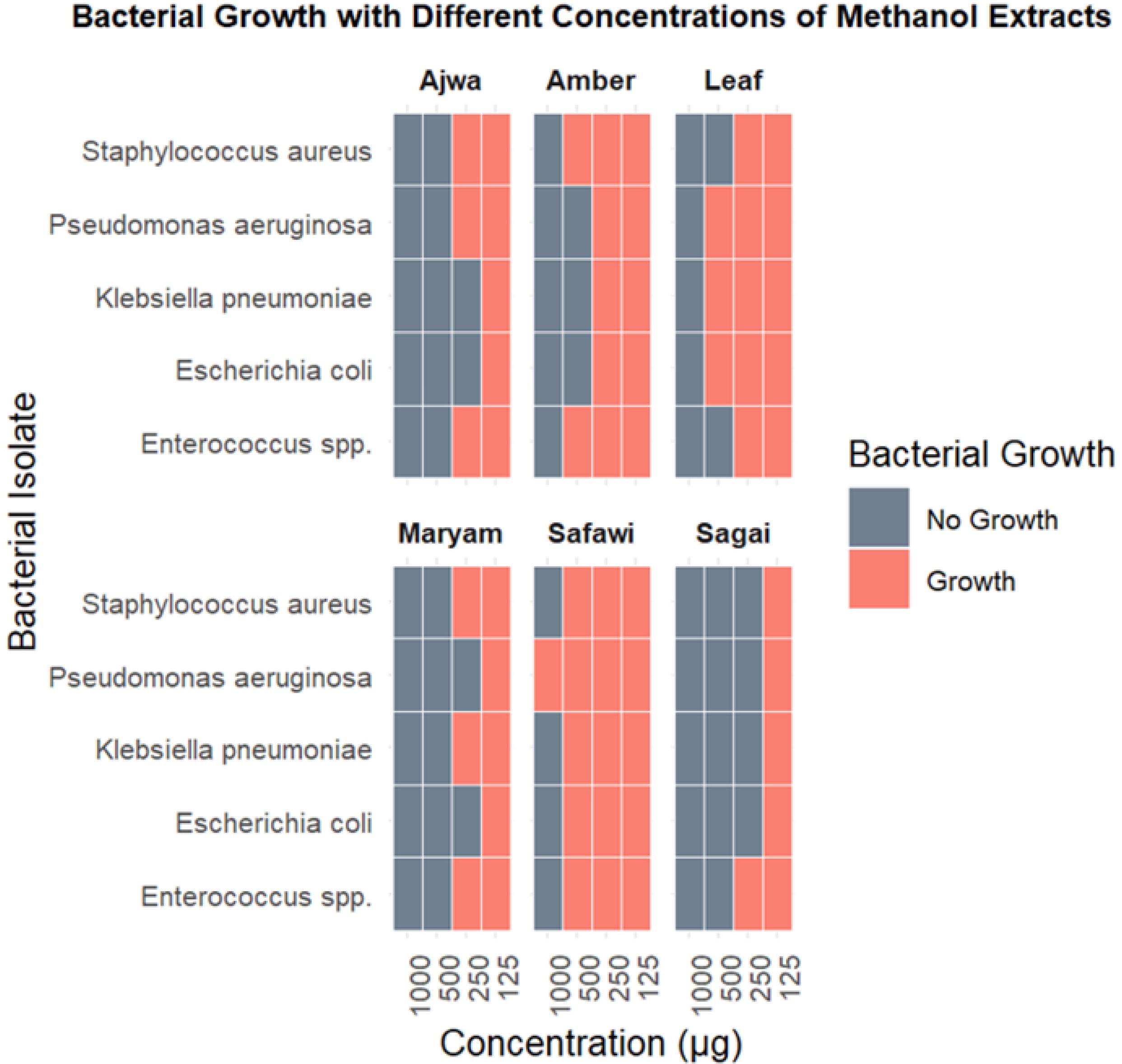
Figure 5.
Bacterial Growth with Different Concentrations of Methanol Extracts of Date Fruits and Leaves
.
Bacterial Growth with Different Concentrations of Methanol Extracts of Date Fruits and Leaves
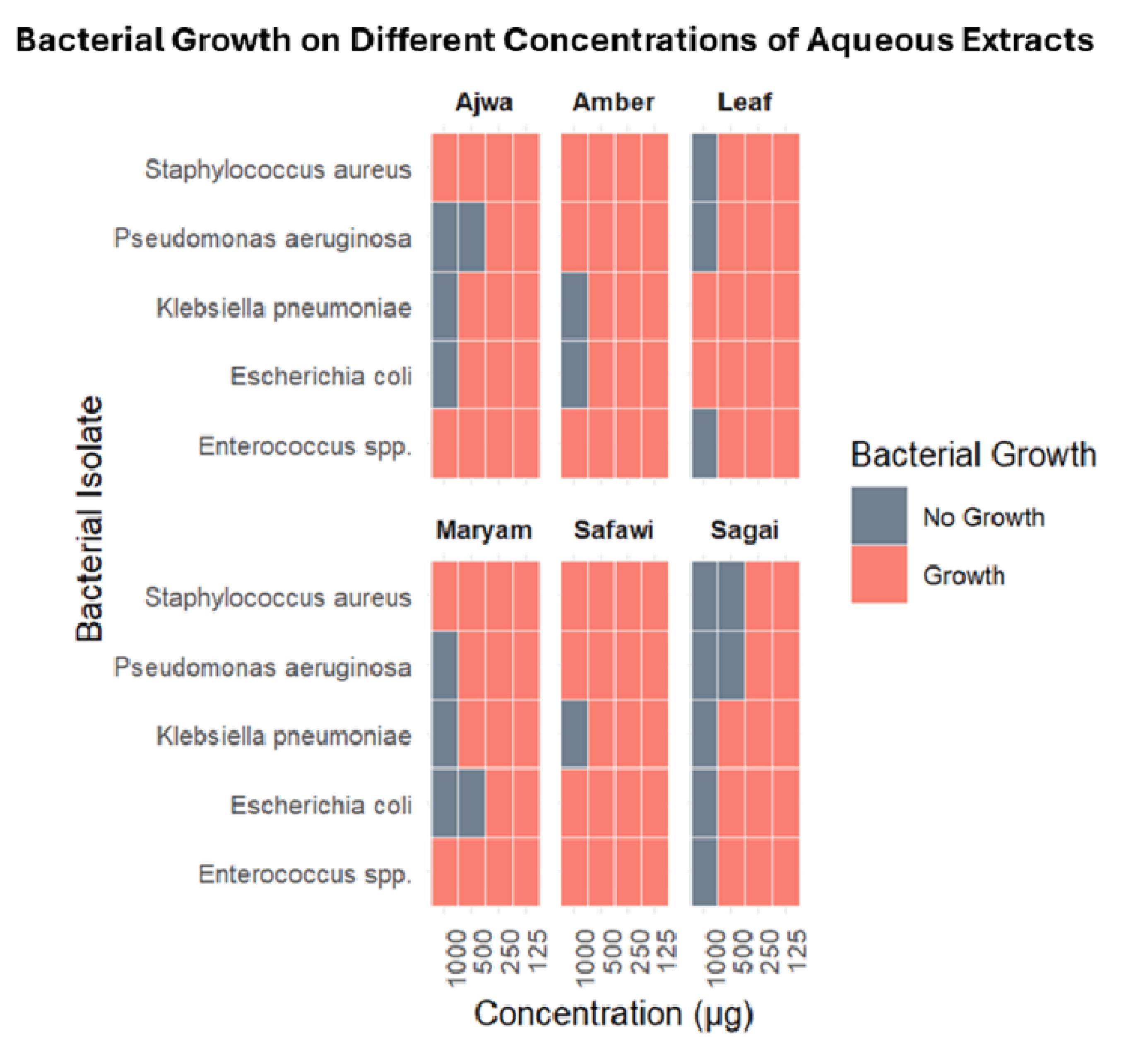
Figure 6.
Bacterial Growth with Different Concentrations of Aqueous Extracts of Date Fruits and Leaves
.
Bacterial Growth with Different Concentrations of Aqueous Extracts of Date Fruits and Leaves
Quantitative MIC assessment confirmed the lowest MIC (187.5 µg/mL) for ethanol extracts of Maryam (E. coli, P. aeruginosa, and Enterococcus spp.) and Sagai (E. coli, K. pneumoniae, and Enterococcus spp.). Comparable MICs were recorded for methanol extracts of Maryam (E. coli and P. aeruginosa), Sagai (all tested bacteria except Enterococcus spp.), and Ajwa (E. coli and K. pneumoniae). In contrast, aqueous extracts generally had MIC values ≥ 375 µg/mL or no detectable inhibition at tested concentrations (Table 2, Figure 7).
Table 2.
Minimum Inhibitory Concentration of Different Extracts (Measured as Means ± SD) Against Five Pathogens (Concentrations in µg/mL)
|
Samples/Isolates
|
Amber
|
Safawi
|
Maryam
|
Sagai
|
Ajwa
|
Leaf
|
|
Ethanol
|
Methanol
|
Aquaous
|
Ethanol
|
Methanol
|
Aquaous
|
Ethanol
|
Methanol
|
Aquaous
|
Ethanol
|
Methanol
|
Aquaous
|
Ethanol
|
Methanol
|
Aquaous
|
Ethanol
|
Methanol
|
Aquaous
|
|
Escherichia coli (n = 3)
|
375 ± 0 |
375 ± 0 |
750 ± 0 |
750 ± 0 |
750 ± 0 |
0 |
187.5 ± 0 |
187.5 ± 0 |
375 ± 0 |
187.5 |
187.5 ± 0 |
750 ± 0 |
375 ± 0 |
187.5 ± 0 |
750 ± 0 |
375 ± 0 |
750 ± 0 |
0 |
|
Significance
|
*** |
*** |
*** |
*** |
*** |
*** |
Pseudomonas aeruginosa
(n = 5)
|
750 ± 0 |
375 |
0 |
750 ± 0 |
0 |
0 |
187.5 ± 0 |
187.5 ± 0 |
750 |
± 0375 |
187.5 ± 0 |
375 ± 0 |
750 ± 0 |
375 ± 0 |
375 ± 0 |
375 ± 0 |
750 ± 0 |
750 ± 0 |
|
Significance
|
*** |
*** |
*** |
*** |
*** |
*** |
|
Staphylococcus aureus (n = 4)
|
375 ± 0 |
750 ± 0 |
0 |
0 |
750 ± 0 |
0 |
375 ± 0 |
375 |
± 00 |
375 ± 0 |
187.5 ± 0 |
375 ± 0 |
750 ± 0 |
375 ± 0 |
0 |
750 ± 0 |
375 ± 0 |
750 ± 0 |
|
Significance
|
*** |
*** |
*** |
*** |
*** |
*** |
Klebsiella pneumoniae
(n = 2)
|
750 ± 0 |
375 ± 0 |
750 ± 0 |
750 ± 0 |
750 ± 0 |
750 ± 0 |
375 ± 0 |
375 ± 0 |
750 ± 0 |
187.5 ± 0 |
187.5 ± 0 |
750 ± 0 |
375 ± 0 |
187.5 ± 0 |
750 ± 0 |
375 ± 0 |
750 ± 0 |
0 |
|
Significance
|
*** |
*** |
*** |
*** |
ns |
*** |
|
Enterococcus spp. (n = 2) |
375 ± 0 |
750 ± 0 |
0 |
0 |
750 |
0 |
187.5 |
375 ± 0 |
0 |
375 ± 0 |
375 ± 0 |
750 ± 0 |
750 ± 0 |
375 ± 0 |
0 |
750 ± 0 |
375 ± 0 |
750 ± 0 |
|
Significance
|
*** |
*** |
*** |
*** |
0.465 (ns) |
*** |
Note. MIC: The lowest concentration of extract inhibiting growth + highest concentration that allows growth. ***P < 0.001.
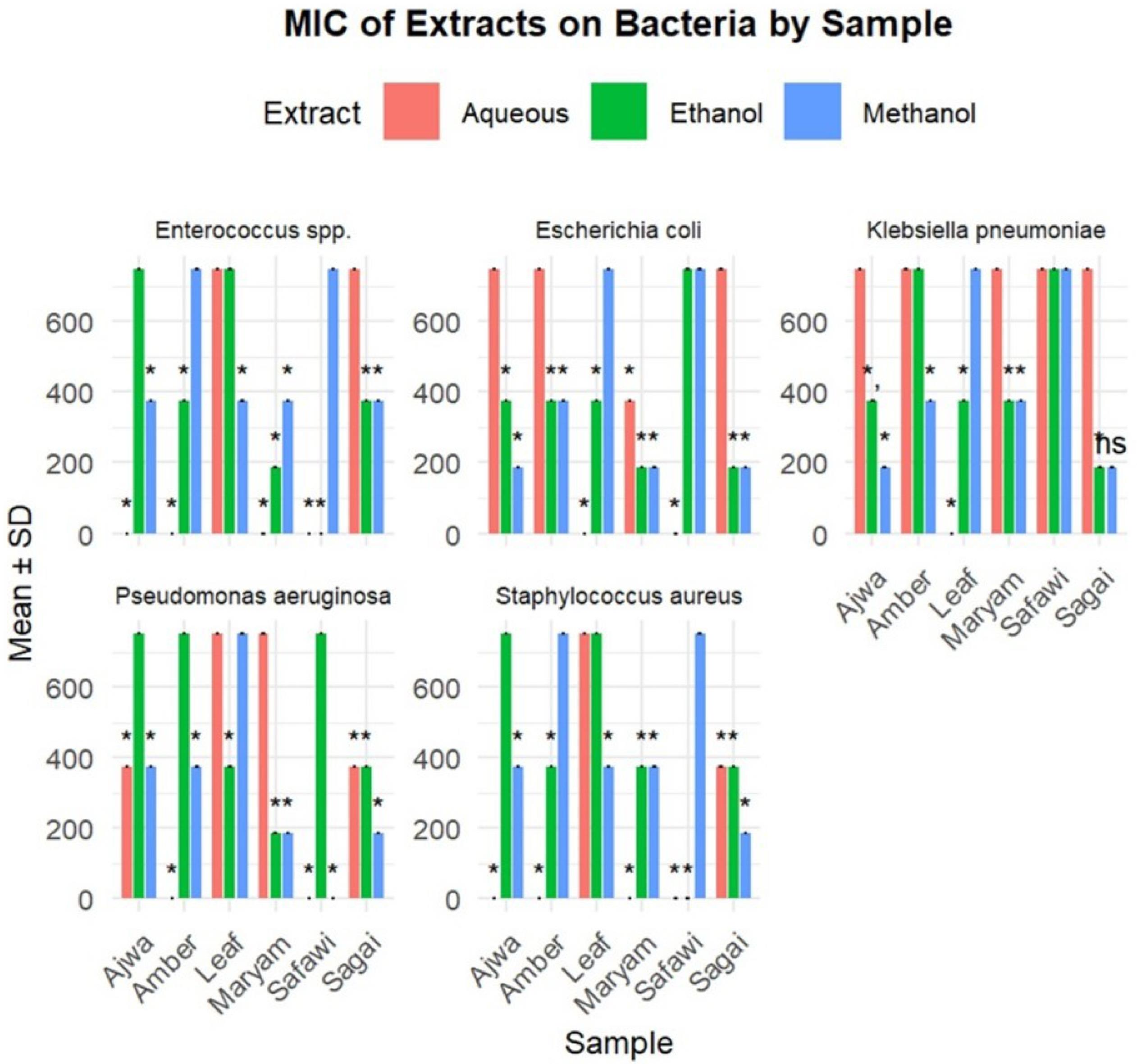
Figure 7.
MIC of Different Extracts on Bacteria. Note. MIC: Minimal inhibitory concentration
.
MIC of Different Extracts on Bacteria. Note. MIC: Minimal inhibitory concentration
Statistical analysis revealed significant differences in MIC values among extract types and bacterial species (P < 0.001), supporting the superior antibacterial efficacy of ethanol and methanol extracts compared to aqueous extracts. K. pneumoniae demonstrated no significant difference between methanol and aqueous extracts (P > 0.05).
Minimum Bactericidal Concentration
Bactericidal activity, measured by MBC, was detected only for ethanol and methanol extracts; aqueous extracts did not exhibit bactericidal effects at the tested concentrations. The ethanol extract of Maryam showed the lowest MBC compared to P. aeruginosa at 350 ± 111.8 µg/mL. Significant differences (P < 0.05) in MBC values were observed for P. aeruginosa between Maryam versus Ajwa, Sagai versus Ajwa, and Maryam versus leaf extracts. For E. coli, S. aureus, K. pneumoniae, and Enterococcus spp., no significant differences in MBC were found across extracts. Maryam, Sagai, and Amber extracts consistently demonstrated lower MBC values, indicating stronger bactericidal activity (Table 3, Figure 8).
Table 3.
Determination of Minimal Bactericidal Concentration of Extracts (Concentrations in µL)
|
Samples/Isolates
|
Amber
|
Safawi
|
Maryam
|
Sagai
|
Ajwa
|
Leaf
|
|
Ethanol
|
Methanol
|
Aquaous
|
Ethanol
|
Methanol
|
Aquaous
|
Ethanol
|
Methanol
|
Aquaous
|
Ethanol
|
Methanol
|
Aquaous
|
Ethanol
|
Methanol
|
Aquaous
|
Ethanol
|
Methanol
|
Aquaous
|
|
Escherichia coli (n = 3)
|
1000 ± 0 |
- |
- |
- |
- |
- |
666.7 ± 288.7 |
666.7 ± 288.7 |
- |
1000 ± 0 |
666.7 ± 288.7 |
- |
- |
666.7 ± 288.7 |
- |
- |
- |
- |
|
Significance
|
Amber vs. Maryam: 0.1876, Amber vs. Sagai: 1.0000, Amber vs. Ajwa: 0.1876, Maryam vs. Sagai: 0.1876, Maryam vs. Ajwa: 1.0000, Sagai vs. Ajwa: 0.1876 |
Pseudomonas aeruginosa
(n = 5)
|
- |
- |
- |
- |
- |
- |
350 ± 111.8 |
700 ± 258.2 |
- |
600 ± 244.9 |
1000 ± 0 |
- |
- |
- |
- |
700 ± 258.2 |
- |
- |
|
Significance
|
Maryam vs. Ajwa: Significant (0.0129), Sagai vs. Ajwa: Significant (0.0339), Maryam vs. Leaf: Significant (0.0413), Maryam vs. Sagai: 0.0558, Ajwa vs. Leaf: 0.1011, Sagai vs. Leaf: 0.6005 |
|
Staphylococcus aureus (n = 4)
|
- |
- |
- |
- |
- |
- |
1000 ± 0 |
1000 ± 0 |
- |
- |
750 ± 288.7 |
- |
- |
- |
- |
1000 ± 0 |
750 ± 288.7 |
- |
|
Significance
|
Maryam vs. Ajwa: 0.1814, Maryam vs. Leaf: 0.1814, Ajwa vs. Leaf: 1.0000 |
Klebsiella pneumoniae
(n = 2)
|
- |
- |
- |
- |
- |
- |
1000 ± 0 |
1000 ± 0 |
- |
- |
500 ± 0 |
- |
- |
750 ± 353.6 |
- |
- |
- |
- |
|
Significance
|
Maryam vs. Ajwa: 0.6171 |
|
Enterococcus spp. (n = 2) |
- |
- |
- |
- |
- |
- |
750 ± 353.6 |
1000 ± 0 |
- |
- |
- |
- |
- |
- |
- |
- |
- |
- |
|
Significance
|
Insufficient data for the test |
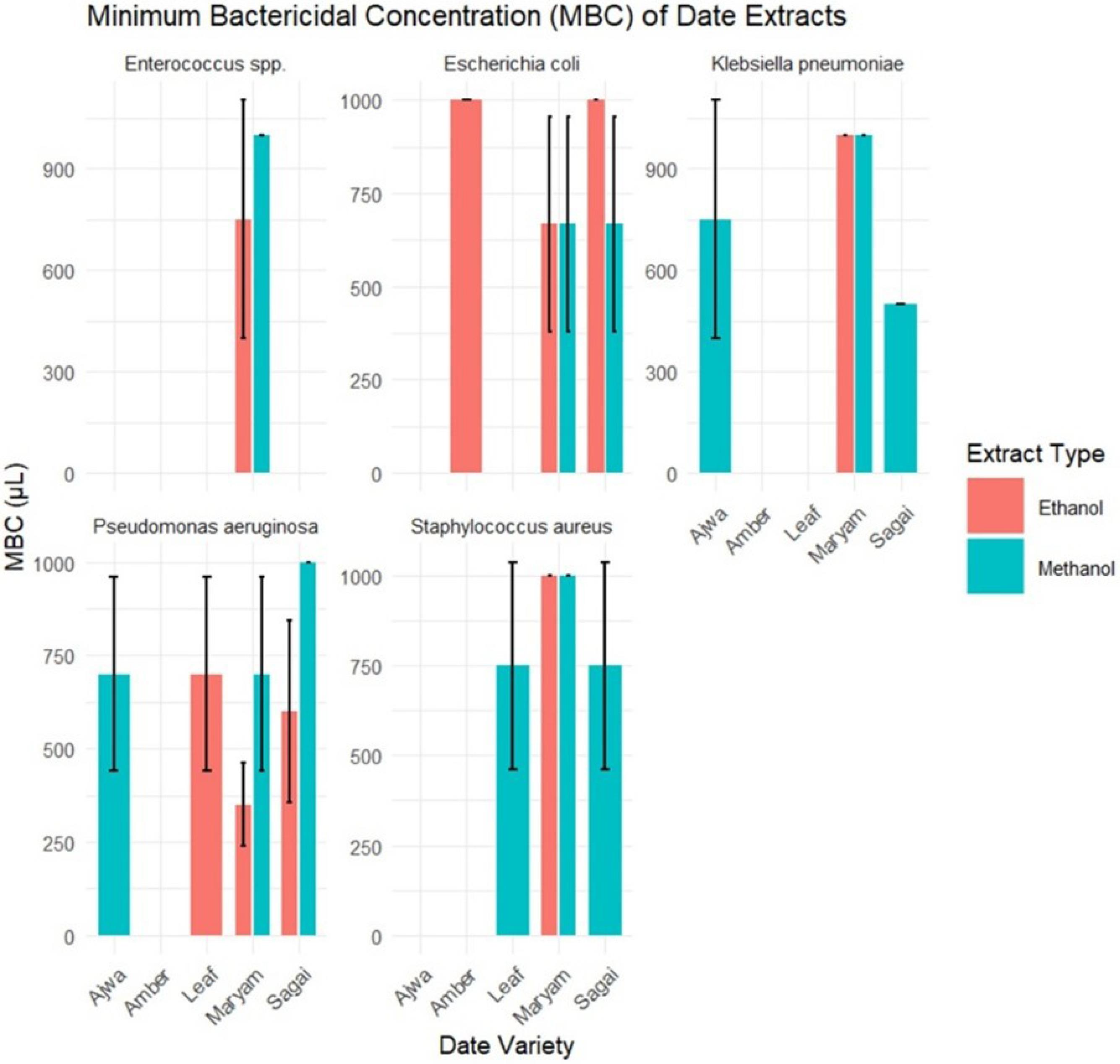
Figure 8.
MBC of Extracts Against Bacteria. Note. MBC: Minimal bactericidal concentration
.
MBC of Extracts Against Bacteria. Note. MBC: Minimal bactericidal concentration
Discussion
To contextualize the novelty of our study, we compiled a comparative summary of previous research on the antimicrobial activity of P. dactylifera (Table 4). Most prior studies focused on fruit or seed extracts, used basic diffusion methods without MIC/MBC quantification, tested a limited range of bacterial species, rarely included multiple varieties, and did not assess leaf extracts or MDR clinical isolates.
Table 4.
Summary of Previous Studies on the Antimicrobial Activity of Phoenix dactylifera Compared to the Present Study
|
Study (Author, Year)
|
Plant Part Used
|
Extract Type/Method
|
Microorganisms Tested
|
MDR Strains
|
MIC/MBC Reported
|
Variety Comparison
|
Notable Gaps/Limitations
|
| Al-Daihan et al, 2012 (27) |
Fruit |
Disc diffusion |
Staphylococcus aureus, Streptococcus pyogenes, Escherichia coli, and Pseudomonas aeruginosa |
No |
No |
✕ |
Only fruit; no MIC/MBC |
| Garba et al, 2013 (28) |
Leaf |
Disc diffusion |
Escherichia coli, Moraxella morganii, Proteus mirabilis, and Yersinia enterocolitica |
No |
Yes (12.5–100 μg/mL) |
✕ |
Limited to leaf only |
| Parveen et al, 2012 (29) |
Leaf, Pit |
Agar well and MIC |
Bacillus subtilis, Escherichia coli, Enterococcus faecalis, Pseudomonas aeruginosa, Shigella flexneri, Staphylococcus aureus, and Streptococcus pyogenes |
No |
Yes |
✓ (3 varieties) |
No MDR strains; unspecified variety details |
| Ayachi et al, 2012 (30) |
Fruit |
Disc–agar diffusion |
Salmonella typhi and Escherichia coli |
No |
No |
✕ |
Few strains; only fruit |
| Yassein et al, 2012 (31) |
Seed |
Agar well diffusion |
Escherichia coli, Klebsiella pneumoniae, Proteus vulgaris, and Pseudomonas aeruginosa |
No |
No |
✕ |
No MIC; inactive against K. pneumoniae |
| Bhat et al, 2012 (32) |
Fruit |
Disc diffusion and MIC |
Staphylococcus aureus, Streptococcus pyogenes, Bacillus subtilis, Escherichia coli, and Pseudomonas aeruginosa |
No |
Yes |
✕ |
Did not compare varieties |
| Shakibaie et al, 2011 (33) |
Seed |
MIC/MBC |
Staphylococcus aureus, Escherichia coli, Bacillus cereus, Salmonella dysenteriae, Salmonella typhi, Klebsiella pneumoniae, Serratia marcescens, and Candida albicans |
No |
Yes (5–40 mg/mL) |
✕ |
K. pneumoniae, S. marcescens, C. albicans inactive |
| Bolin et al, 1972 (34) |
Fruit |
Storage extract |
Pseudomonas aeruginosa, Enterococcus faecalis, Diphtheroid coryneform bacteria, Proteus vulgaris, and Escherichia coli |
No |
No |
✕ |
Old study; moisture-controlled whole fruit |
| Sallal et al, 2013 (35) |
Fruit |
Agar well |
Staphylococcus aureus
|
No |
No |
✕ |
One strain only |
| Mahmood et al, 2012 (36) |
Fruit |
Disc diffusion |
Escherichia coli, Staphylococcus aureus, and Pseudomonas aeruginosa |
No |
No |
✕ |
Basic diffusion; no MIC |
| Al-Seeni et al, 2012 (37) |
Fruit |
Disc–agar diffusion |
Pseudomonas aeruginosa, Escherichia coli, Shigella, Klebsiella pneumoniae, Bacillus subtilis, Staphylococcus aureus, and Micrococcus |
No |
No |
✕ |
No quantification of potency |
| Selim et al, 2014 (38) |
Fruit |
Disc diffusion |
Pseudomonas aeruginosa
|
No |
No |
✕ |
Single organism tested |
| Amiour et al, 2014 (39) |
Fruit |
Disc diffusion |
Staphylococcus aureus, Pseudomonas aeruginosa, Klebsiella pneumoniae, Escherichia coli, Bacillus subtilis, and Staphylococcus spp. |
No |
No |
✕ |
No MIC/MBC |
| Abu-Elteen et al, 2000 (40) |
Fruit |
Antifungal and MIC |
Candida albicans
|
No |
Yes |
✕ |
Only fungal strains tested |
| Sallal et al, 1996 (41) |
Fruit |
MIC and germ tube |
Candida albicans
|
No |
Yes |
✕ |
Fungal only |
| Sharideh et al, 1998 (42) |
Fruit |
MIC, morphology |
Candida albicans
|
No |
Yes |
✕ |
Fungal only |
| Current study (2025) |
Fruit + Leaf |
Ethanol, methanol, water, and crude |
Staphylococcus aureus, Klebsiella pneumoniae, Escherichia coli, and Pseudomonas aeruginosa (MDR clinical isolates) |
✓ |
✓ |
✓ (5 varieties) |
First to compare 5 named varieties + leaf extracts against MDR clinical isolates |
Note. MDR: Multidrug-resistant; MIC: Minimal inhibitory concentration; MBC: Minimal bactericidal concentration.
In contrast, our study is the first to evaluate five named varieties of P. dactylifera along with their leaf extracts, using aqueous crude homogenates and solvent-based extractions against the MDR clinical isolates of S. aureus, E. coli, K. pneumoniae, P. aeruginosa, and Enterococcus spp., reporting quantitative MIC and MBC values. These pathogens cause serious infections and exhibit increasing resistance due to factors such as unregulated antibiotic use (43-50).
Our antimicrobial susceptibility tests confirmed high resistance, particularly in P. aeruginosa and Enterococcus spp. Ethanol and methanol extracts demonstrated stronger antibacterial activity than aqueous extracts, likely due to the improved extraction of phenolics and flavonoids. Among varieties, Maryam, Sagai, and Amber showed potent activity, with MICs as low as 187.5 µg/mL and significant bactericidal effects—especially Maryam ethanol extract against P. aeruginosa (MBC 350 ± 111.8 µL). The Safawi methanol extract was most effective against K. pneumoniae. Statistical analyses highlighted the influence of solvent and variety on efficacy.
Our findings are consistent with those of earlier studies, demonstrating the antibacterial properties of date extracts (51) and further advancing this understanding by including MDR pathogens and leaf-derived extracts. The observed bactericidal concentrations indicate therapeutic promise for certain varieties.
Varietal differences in antibacterial activity are likely due to differences in phytochemical composition, which are influenced by cultivar genetics, ripening stage, and geographical factors, such as climate, soil, and water availability (52). Prior research identified flavonoids, tannins, phenolic acids, and polyphenols in date fruits and leaves, compounds known to disrupt bacterial membranes, inhibit nucleic acid synthesis, and interfere with metabolic enzymes (53,54). Due to equipment limitations, it was impossible to conduct phytochemical profiling in this study, but the presence of these bioactives is well documented in the literature (52-54).
The enhanced activity of ethanol and methanol extracts over aqueous ones can be attributed to the higher solubility of phenolic compounds in organic solvents (55). This is in line with the findings of previous research, showing that organic solvents improve the extraction of active constituents, such as flavonoids and phenolic acids (55).
Compounds such as quercetin and catechin—abundant in dates—are known to damage bacterial membranes and promote the formation of reactive oxygen species, which play a critical role in killing Gram-negative bacteria (56,57). These compounds also inhibit bacterial DNA replication and protein synthesis (57).
Tannins, also present in date varieties, may exert additional effects by precipitating microbial proteins, inactivating enzymes, and reducing virulence, including adherence and biofilm formation (58).
The difference in antimicrobial efficacy across varieties further suggests the role of specific phytochemical profiles, which vary by variety and region (59). For example, differences in flavonoid or tannin content between Amber and Safawi could account for their varied MIC/MBC values.
Moreover, potential synergistic effects among different phytochemicals—such as phenolic–flavonoid or tannin–flavonoid interactions—may enhance antibacterial efficacy, especially against MDR bacteria (60). This warrants further investigation through fractionation and bioassay-guided studies.
The demonstrated antibacterial activity of P. dactylifera extracts, particularly those derived using ethanol and methanol, suggests potential for therapeutic application, especially in the context of MDR infections. Given their observed efficacy against skin-associated and wound-associated pathogens, such as S. aureus and P. aeruginosa, these extracts may be explored for topical use in the form of ointments or wound dressings (61). However, systemic application would require further investigation into the extracts’ toxicity, bioavailability, and pharmacokinetics (62). The natural origin of these compounds offers appeal for developing plant-based adjuncts or alternatives to conventional antibiotics, particularly in resource-limited settings where resistance is prevalent and access to advanced treatments is restricted.
In summary, ethanol and methanol extracts of P. dactylifera varieties exhibit promising antibacterial and bactericidal activity against MDR pathogens, likely driven by phenolic compounds and influenced by varietal phytochemistry. Future research should include phytochemical analyses (e.g., thin-layer chromatography, high-performance liquid chromatography, and UV-Vis spectroscopy) to identify active constituents and clarify their mechanisms. Understanding synergistic interactions and varietal bioactivity differences will be critical in developing date-based antimicrobial agents.
Limitations of the Study
A critical limitation of this study was the absence of detailed phytochemical profiling techniques, such as thin-layer chromatography, high-performance liquid chromatography, or UV-Vis spectroscopic analyses, which were not accessible in our laboratory during this research. This absence significantly limited our ability to identify and quantify the specific bioactive compounds responsible for the observed antibacterial activity. Consequently, attributing the antibacterial effects to phenolics, flavonoids, tannins, or other phytochemicals remains speculative and unconfirmed.
Another limitation was the reliance on clinical isolates rather than ATCC reference strains, which may impact the reproducibility of our results. The unavailability of ATCC reference strains in our laboratory was a constraint for this study. Future work should incorporate these reference strains to validate the antibacterial activity and ensure greater reproducibility of the findings.
While our primary objective was to establish whether extracts from dates and their leaves exhibit antibacterial activity against pathogenic isolates, we recognize that mechanistic insights into which compounds drive this activity are essential to fully understand and validate these effects. Therefore, the lack of phytochemical characterization and the absence of ATCC strains represent significant gaps in the current work.
It is strongly recommended that future research prioritize comprehensive phytochemical analyses to isolate, identify, and quantify the key antimicrobial constituents. Such analyses will not only confirm the roles of phenolic and flavonoid compounds but also help elucidate their mechanisms of action, particularly against MDR bacterial strains. Similarly, the inclusion of ATCC reference strains in future studies will enhance reproducibility and the robustness of antibacterial efficacy results.
Conclusion
Natural products are promising alternatives in the search for antibacterial agents effective against antibiotic-resistant bacteria. Numerous plants, fruits, leaves, and barks have demonstrated potential in infection treatment. Dates and their leaves, in particular, demonstrated antibacterial activity against several MDR clinical isolates. The next step involves identifying the specific phytochemicals responsible, which could pave the way for developing these compounds as therapeutic agents.
Acknowledgements
The authors are thankful to the Department of Microbiology, Stamford University, Bangladesh, for their logistics and laboratory support during the research.
Competing Interests
The authors declare that they have no conflict of interests.
Ethical Approval
Ethical approval was not required for this study, as no human participants or personal data were involved. The bacterial isolates used in this study were archived strains originally preserved in the teaching laboratory of the Department of Microbiology for undergraduate practical classes and student thesis projects. These isolates were utilized solely for laboratory-based antimicrobial testing, with no access to clinical records or patient-identifiable information. Their use complies with the institutional policy for educational and research use of anonymized microbial cultures.
Funding
The cost for the materials and reagents was covered by the Department of Microbiology, Stamford University, Bangladesh.
References
- Jaganathan V, Shanmugavadivu M, Ganesh S. Preliminary phytochemical screening and anti-bacterial activity of date seed methanolic extract. Int J Adv Res Biol Sci 2018; 5(2):209-15. doi: 10.22192/ijarbs.2018.05.02.021 [Crossref] [ Google Scholar]
- Al-Orf SM, Ahmed MH, Al-Atwai N, Al-Zaidi H, Dehwah A, Dehwah S. Nutritional properties and benefits of the date fruits (Phoenix dactylifera L). Bull Natl Nutr Inst Arab Repub Egypt 2012; 39:97-129. [ Google Scholar]
- Al-Alawi RA, Al-Mashiqri JH, Al-Nadabi JS, Al-Shihi BI, Baqi Y. Date palm tree (Phoenix dactylifera L): natural products and therapeutic options. Front Plant Sci 2017; 8:845. doi: 10.3389/fpls.2017.00845 [Crossref] [ Google Scholar]
- Shariati M, Sharifi E, Kaveh M. The effect of Phoenix dactylifera (date-palm) pit powder on testosterone level and germ cells in adult male rats. J Adv Med Biomed Res 2007;15(61):21-7. [Persian].
- Alghamdi AA, Awadelkarem AM, Hossain AB, Ibrahim NA, Fawzi M, Ashraf SA. Nutritional assessment of different date fruits (Phoenix dactylifera L) varieties cultivated in Hail province, Saudi Arabia. Biosci Biotechnol Res Commun 2018; 11(2):263-9. doi: 10.21786/bbrc/11.1/11 [Crossref] [ Google Scholar]
- Khalid S, Ahmad A, Masud T, Asad MJ, Sandhu M. Nutritional assessment of Ajwa date flesh and pits in comparison to local varieties. J Anim Plant Sci 2016; 26(4):1072-80. [ Google Scholar]
- Rauha JP, Remes S, Heinonen M, Hopia A, Kähkönen M, Kujala T. Antimicrobial effects of Finnish plant extracts containing flavonoids and other phenolic compounds. Int J Food Microbiol 2000; 56(1):3-12. doi: 10.1016/s0168-1605(00)00218-x [Crossref] [ Google Scholar]
- Puupponen-Pimiä R, Nohynek L, Meier C, Kähkönen M, Heinonen M, Hopia A. Antimicrobial properties of phenolic compounds from berries. J Appl Microbiol 2001; 90(4):494-507. doi: 10.1046/j.1365-2672.2001.01271.x [Crossref] [ Google Scholar]
- Zhu X, Zhang H, Lo R. Phenolic compounds from the leaf extract of artichoke (Cynara scolymus L) and their antimicrobial activities. J Agric Food Chem 2004; 52(24):7272-8. doi: 10.1021/jf0490192 [Crossref] [ Google Scholar]
- Proestos C, Chorianopoulos N, Nychas GJ, Komaitis M. RP-HPLC analysis of the phenolic compounds of plant extracts investigation of their antioxidant capacity and antimicrobial activity. J Agric Food Chem 2005; 53(4):1190-5. doi: 10.1021/jf040083t [Crossref] [ Google Scholar]
- Sousa A, Ferreira IC, Calhelha R, Andrade PB, Valentão P, Seabra R. Phenolics and antimicrobial activity of traditional stoned table olives ‘alcaparra’. Bioorg Med Chem 2006; 14(24):8533-8. doi: 10.1016/j.bmc.2006.08.027 [Crossref] [ Google Scholar]
- Pereira JA, Pereira AP, Ferreira IC, Valentão P, Andrade PB, Seabra R. Table olives from Portugal: phenolic compounds, antioxidant potential, and antimicrobial activity. J Agric Food Chem 2006; 54(22):8425-31. doi: 10.1021/jf061769j [Crossref] [ Google Scholar]
- Negi PS, Jayaprakasha GK. Antioxidant and antibacterial activities of Punica granatum peel extracts. J Food Sci 2003; 68(4):1473-7. doi: 10.1111/j.1365-2621.2003.tb09669.x [Crossref] [ Google Scholar]
- Shen X, Sun X, Xie Q, Liu H, Zhao Y, Pan Y. Antimicrobial effect of blueberry (Vaccinium corymbosum L) extracts against the growth of Listeria monocytogenes and Salmonella Enteritidis. Food Control 2014; 35(1):159-65. doi: 10.1016/j.foodcont.2013.06.040 [Crossref] [ Google Scholar]
- Mucha P, Skoczyńska A, Małecka M, Hikisz P, Budzisz E. Overview of the Antioxidant and Anti-Inflammatory Activities of Selected Plant Compounds and Their Metal Ions Complexes. Molecules 2021;26(16). doi: 10.3390/molecules26164886.
- Gu L, Kelm MA, Hammerstone JF, Beecher G, Holden J, Haytowitz D. Screening of foods containing proanthocyanidins and their structural characterization using LC-MS/MS and thiolytic degradation. J Agric Food Chem 2003; 51(25):7513-21. doi: 10.1021/jf034815d [Crossref] [ Google Scholar]
- Khalid S, Ahmad A, Kaleem M. Antioxidant activity and phenolic contents of Ajwa date and their effect on lipo-protein profile. Funct Food Health Dis 2017; 7(6):396-410. doi: 10.31989/ffhd.v7i6.337 [Crossref] [ Google Scholar]
- Khalid S, Khalid N, Khan RS, Ahmed H, Ahmad A. A review on chemistry and pharmacology of Ajwa date fruit and pit. Trends Food Sci Technol 2017; 63:60-9. doi: 10.1016/j.tifs.2017.02.009 [Crossref] [ Google Scholar]
- Isaka I, Gumel AM, Muhammad HU, Kemi AF. Phytochemical analysis and antimicrobial activity of Neocarya macrophylla leaves extract. Int J Health Life Sci 2017; 3(1):18-34. doi: 10.20319/lijhls.2017.31.1834 [Crossref] [ Google Scholar]
- Davidson PM. Chemical preservatives and natural antimicrobial compounds. In: Doyle MP, Beuchat LR, Montville TJ, eds. Food Microbiology: Fundamentals and Frontiers. 2nd ed. Washington, DC: ASM Press; 2001. p. 593-627.
- Abdul-Hamid NA, Abas F, Ismail IS, Shaari K, Lajis NH. Influence of different drying treatments and extraction solvents on the metabolite profile and nitric oxide inhibitory activity of Ajwa dates. J Food Sci 2015; 80(11):H2603-11. doi: 10.1111/1750-3841.13084 [Crossref] [ Google Scholar]
- Taleb H, Maddocks SE, Morris RK, Kanekanian AD. Chemical characterisation and the anti-inflammatory, anti-angiogenic and antibacterial properties of date fruit (Phoenix dactylifera L). J Ethnopharmacol 2016; 194:457-68. doi: 10.1016/j.jep.2016.10.032 [Crossref] [ Google Scholar]
- Ferraro MJ, Craig WA, Dudley MN. Performance Standards for Antimicrobial Susceptibility Testing. 11th ed. Wayne, PA: NCCLS; 2001.
- Clinical and Laboratory Standards Institute (CLSI). CLSI Document M07-A9. Methods for Dilution Antimicrobial Susceptibility Tests for Bacteria that Grow Aerobically: Approved Standard. 9th ed. Wayne, PA: CLSI; 2012.
- Manokari SL, Meenu NC. A study on the extraction process of Wrightia tinctoria and evaluation of its antimicrobial activity. Int J Res Appl Sci Eng Technol 2017; 5(9):1308-16. doi: 10.22214/ijraset.2017.9188 [Crossref] [ Google Scholar]
- Jorgensen JH, Turnide JD, Washington JA. Antibacterial susceptibility tests: dilution and disk diffusion method. In: Murray PR, Pfaller MA, Tenover FC, Baron EJ, Yolken RH, eds. Manual of Clinical Microbiology. 7th ed. Washington, DC: ASM Press; 1999. p. 1526-43.
- Al-Daihan S, Bhat RS. Antibacterial activities of extracts of leaf, fruit, seed and bark of Phoenix dactylifera. Afr J Biotechnol 2012; 11(42):10021-5. doi: 10.5897/ajb11.4309 [Crossref] [ Google Scholar]
- Garba IH, Shehu K, Salihu L. Antibacterial activity of date palm (Phoenix dactylifera) leaf extracts against some selected bacterial pathogens. Bayero J Pure Appl Sci 2013; 6:96-100. [ Google Scholar]
- Perveen K, Bokhari NA, Shah AH. Antibacterial activity of Phoenix dactylifera L leaves and pits extracts against selected bacterial pathogens. J Med Plants Res 2012; 6:3725-9. [ Google Scholar]
- Ayachi A, Alloui N, Bennoune O, Alloui MN. Antibacterial activity of some fruits: preliminary results. Vet World 2009; 2:410-2. [ Google Scholar]
- Yassein SA. Antibacterial activity of some extracts from date palm seeds (Phoenix dactylifera L) against multidrug resistant bacteria. Iraqi J Sci 2012; 53:561-7. [ Google Scholar]
- Bhat RS, Al-Daihan S. Phytochemical constituents and antibacterial activity of some green leafy vegetables. Asian Pac J Trop Biomed 2012; 2:S1643-6. [ Google Scholar]
- Shakibaie M, Salari MH, Mosavi HA. Antibacterial and antifungal effects of date palm pit (seed) extract. Jundishapur J Microbiol 2011; 4:183-7. [ Google Scholar]
- Bolin HR, Stafford AE, King AD. Antimicrobial activity of dehydrated dates. Appl Microbiol 1972; 23:938-43. [ Google Scholar]
- Sallal AK, Hassan MS, Al-Jubori SS. Antibacterial activity of date palm (Phoenix dactylifera L) pit extract against Staphylococcus aureus. Iraqi J Sci 2013; 54:667-70. [ Google Scholar]
- Mahmood A, Abdul Ghani B, Ikram U. Evaluation of antimicrobial activity of date extracts against gram-positive and gram-negative bacteria. Afr J Microbiol Res 2012; 6:3812-5. [ Google Scholar]
- Al-Seeni MN. Evaluation of antibacterial activity of date palm fruit extracts. Int J Pharm Sci Res 2012; 3:4335-40. [ Google Scholar]
- Selim SA, Al Jaouni SK, Deraz SF, Othman MA. Antimicrobial activity of essential oils isolated from medicinal plants against multidrug-resistant bacteria. Saudi Med J 2014; 35:437-46. [ Google Scholar]
- Amiour N, Boucherit K, Boucherit-Otmani Z, Abdi A, Kechrid A. Antibacterial activity of date palm (Phoenix dactylifera L) fruits extracts from Algeria against clinical strains. J Chem Pharm Res 2014; 6:78-82. [ Google Scholar]
- Abu-Elteen KH. Antifungal activity of aqueous and methanolic extracts of some date palm (Phoenix dactylifera L) cultivars. Microbios 2000; 103:39-46. [ Google Scholar]
- Sallal AK, Mohammed NA, Naji S. The antifungal activity of date palm (Phoenix dactylifera L) extracts against Candida albicans. Microbios 1996; 86:85-95. [ Google Scholar]
- Shraideh Z, Abu-Elteen KH, Sallal AK. Inhibition of germ tube formation in Candida albicans by aqueous extracts of date palm (Phoenix dactylifera L) pits and flesh. Microbios 1998; 93:35-43. [ Google Scholar]
- Justice SS, Hung C, Theriot JA, Fletcher DA, Anderson GG, Footer MJ. Differentiation and developmental pathways of uropathogenic Escherichia coli in urinary tract pathogenesis. Proc Natl Acad Sci U S A 2004; 101(5):1333-8. doi: 10.1073/pnas.0308125100 [Crossref] [ Google Scholar]
- Driscoll JA, Brody SL, Kollef MH. The epidemiology, pathogenesis and treatment of Pseudomonas aeruginosa infections. Drugs 2007; 67(3):351-68. doi: 10.2165/00003495-200767030-00003 [Crossref] [ Google Scholar]
- Keynan Y, Rubinstein E. The changing face of Klebsiella pneumoniae infections in the community. Int J Antimicrob Agents 2007; 30(5):385-9. doi: 10.1016/j.ijantimicag.2007.06.019 [Crossref] [ Google Scholar]
- Tong SY, Davis JS, Eichenberger E, Holland TL, Fowler VG Jr. Staphylococcus aureus infections: epidemiology, pathophysiology, clinical manifestations, and management. Clin Microbiol Rev 2015; 28(3):603-61. doi: 10.1128/cmr.00134-14 [Crossref] [ Google Scholar]
- Moellering RC Jr. Emergence of Enterococcus as a significant pathogen. Clin Infect Dis 1992; 14(6):1173-6. doi: 10.1093/clinids/14.6.1173 [Crossref] [ Google Scholar]
- Singh SB, Barrett JF. Empirical antibacterial drug discovery--foundation in natural products. Biochem Pharmacol 2006; 71(7):1006-15. doi: 10.1016/j.bcp.2005.12.016 [Crossref] [ Google Scholar]
- Davies J. Resistance redux. EMBO Rep 2007; 8:616-21. [ Google Scholar]
- McKenna M. Antibiotic resistance: the last resort. Nature 2013; 499(7459):394-6. doi: 10.1038/499394a [Crossref] [ Google Scholar]
- Perveen K, Bokhari NA, Soliman DA. Antibacterial activity of Phoenix dactylifera L leaf and pit extracts against selected gram-negative and gram-positive pathogenic bacteria. J Med Plants Res 2012; 6(2):296-300. doi: 10.5897/jmpr11.1380 [Crossref] [ Google Scholar]
- Al-Farsi M, Alasalvar C, Morris A, Baron M, Shahidi F. Compositional and sensory characteristics of three native sun-dried date (Phoenix dactylifera L) varieties grown in Oman. J Agric Food Chem 2005; 53(19):7586-91. doi: 10.1021/jf050578y [Crossref] [ Google Scholar]
- Mohamed EA, Muddathir AM, Osman MA. Antimicrobial activity, phytochemical screening of crude extracts, and essential oils constituents of two Pulicaria spp growing in Sudan. Sci Rep 2020; 10(1):17148. doi: 10.1038/s41598-020-74262-y [Crossref] [ Google Scholar]
- Cushnie TP, Lamb AJ. Antimicrobial activity of flavonoids. Int J Antimicrob Agents 2005; 26(5):343-56. doi: 10.1016/j.ijantimicag.2005.09.002 [Crossref] [ Google Scholar]
- Scalbert A. Antimicrobial properties of tannins. Phytochemistry 1991; 30(12):3875-83. doi: 10.1016/0031-9422(91)83426-l [Crossref] [ Google Scholar]
- Hussain MI, Farooq M, Syed QA. Nutritional and biological characteristics of the date palm fruit (Phoenix dactylifera L) – a review. Food Biosci 2020; 34:100509. doi: 10.1016/j.fbio.2019.100509 [Crossref] [ Google Scholar]
- Borges A, Ferreira C, Saavedra MJ, Simões M. Antibacterial activity and mode of action of ferulic and gallic acids against pathogenic bacteria. Microb Drug Resist 2013; 19(4):256-65. doi: 10.1089/mdr.2012.0244 [Crossref] [ Google Scholar]
- Cowan MM. Plant products as antimicrobial agents. Clin Microbiol Rev 1999; 12(4):564-82. doi: 10.1128/cmr.12.4.564 [Crossref] [ Google Scholar]
- Al-Mssallem MQ, Alqurashi RM, Al-Khayri JM. Bioactive compounds of date palm (Phoenix dactylifera L.). In: Murthy HN, Bapat VA, eds. Bioactive Compounds in Underutilized Fruits and Nuts. Cham: Springer; 2019. p. 1-20.
- Hemaiswarya S, Kruthiventi AK, Doble M. Synergism between natural products and antibiotics against infectious diseases. Phytomedicine 2008; 15(8):639-52. doi: 10.1016/j.phymed.2008.06.008 [Crossref] [ Google Scholar]
- Church D, Elsayed S, Reid O, Winston B, Lindsay R. Burn wound infections. Clin Microbiol Rev 2006; 19(2):403-34. doi: 10.1128/cmr.19.2.403-434.2006 [Crossref] [ Google Scholar]
- Sharma A, Bajpai M. Plant-derived antimicrobial agents: a promising alternative to synthetic antibiotics. Front Pharmacol 2021; 12:698149. [ Google Scholar]