Avicenna Journal of Clinical Microbiology and Infection. 12(2):73-80.
doi: 10.34172/ajcmi.3625
Original Article
Experimental Evaluation of Antibacterial and Antibiofilm Activity of Camellia sinensis, Fraxinus excelsior, and Green Coffee Extracts Against Pseudomonas aeruginosa
Sina Ahmadi Conceptualization, Data curation, Methodology, Project administration, Supervision, 1 
Fatemeh Riyahi Zaniani Conceptualization, Formal analysis, Funding acquisition, Project administration, Supervision, Validation, Writing – original draft, 2, 3, *
Bahar Hasanpour Conceptualization, Data curation, Methodology, Project administration, Supervision, 4
Marzieh An’aam Data curation, Methodology, 4
Author information:
1Student Research Committee, Dezful University of Medical Sciences, Dezful, Iran
2Infectious and Tropical Diseases Research Center, Dezful University of Medical Sciences, Dezful, Iran
3Department of Immunology and Microbiology, School of Medicine, Dezful University of Medical Sciences, Dezful, Iran
4Student Research Committee, Dezful University of Medical Sciences, Dezful, Iran
Abstract
Background: The excessive use of antibiotics has contributed to the emergence of antibiotic-resistant microbes, posing a significant global health concern. Additionally, the formation of biofilms by microorganisms on surfaces further exacerbates the problem by enhancing their resistance to antibacterial agents. Exploring alternative antimicrobial and antibiofilm agents that do not promote drug resistance is crucial to address these issues. This laboratory-based research investigated the antibacterial and antibiofilm efficacy of both water-based and alcohol-based extracts from three medicinal plants, namely, Camellia sinensis, Fraxinus excelsior, and green coffee, against Pseudomonas aeruginosa ATCC 27853 and an extensively drug-resistant (XDR) P. aeruginosa clinical isolate.
Methods: Four distinct methodologies were employed to assess the antibacterial efficacy of the plant extracts, including spot assay, disc diffusion test, agar well diffusion technique, and the micro-broth dilution method. The antibiofilm potential was evaluated using the microtiter plate technique at the sub-inhibitory concentrations of each extract, only against P. aeruginosa ATCC 27853.
Results: The findings revealed that C. sinensis extracts (both aqueous and ethanolic) were the most effective antimicrobials, displaying the lowest minimum inhibitory concentration (MIC) and minimum bactericidal concentration values. While F. excelsior exhibited intermediate antibacterial effects, the green coffee extract lacked substantial antimicrobial action. Furthermore, none of the plant extracts significantly inhibited biofilm formation by P. aeruginosa ATCC 27853.
Conclusion: In general, C. sinensis demonstrated significant potential as an antibacterial agent and showed promising in vitro efficacy. However, the absence of significant biofilm inhibition and high MIC values for other extracts highlight the need for further formulation and mechanistic studies.
Keywords: Camellia sinensis, Fraxinus, Pseudomonas aeruginosa, Antibacterial, Antibiofilm, Drug resistance
Copyright and License Information
© 2025 The Author(s); Published by Hamadan University of Medical Sciences.
This is an open-access article distributed under the terms of the Creative Commons Attribution License (
https://creativecommons.org/licenses/by/4.0), which permits unrestricted use, distribution, and reproduction in any medium, provided the original work is properly cited.
Please cite this article as follows: Ahmadi S, Riyahi Zaniani F, Hasanpour B, An’aam M. Experimental evaluation of antibacterial and antibiofilm activity of Camellia sinensis, Fraxinus excelsior, and green coffee extracts against Pseudomonas aeruginosa. Avicenna J Clin Microbiol Infect. 2025;12(2):73-80. doi:10.34172/ajcmi.3625
Introduction
Increasing antibiotic use causes microorganisms to develop antibiotic resistance, thereby posing a major health challenge worldwide. Multidrug-resistant pathogens now represent a critical threat in both nosocomial and community-related infections (1). Another problem is the irreversible attachment of microorganisms to surfaces, known as biofilms.
Bacterial biofilms form a structured extracellular matrix that protects embedded cells from both host immune responses and antimicrobial agents. This structural barrier contributes to treatment resistance and chronic infection persistence. Recent studies have shown that bacteria within biofilms exhibit intrinsic tolerance to antibiotics, making biofilm-associated infections particularly difficult to eradicate (2). Moreover, Xu et al demonstrated that even advanced antimicrobial strategies, such as photocatalytic oxidation, face limitations in penetrating the biofilm matrix, thus highlighting its critical role in therapeutic failure (3). Therefore, developing antimicrobial and antibiofilm agents without promoting drug resistance should be a priority. Herbal medicine has recently gained popularity due to its non-adverse effects and herbal properties. Compounds such as flavonoids and phenolics found in medicinal plants possess various biological activities, including antibacterial, antioxidative, anti-inflammatory, and anticancer effects (4). Recent reviews have emphasized the potential of phytochemical-based nanomaterials in combating antibiotic-resistant bacteria, emphasizing advancements in nanotechnology applications and the antimicrobial efficacy of flavonoids. These studies underscore the importance of exploring plant-derived compounds as alternative antimicrobial agents (5).
Camellia sinensis, commonly known as tea, is a widely consumed beverage worldwide and contains polyphenolic compounds, such as flavanols, flavonoids, and phenolic acids. Prior studies have reported its efficacy against numerous pathogens (6,7). Coffee is another popular beverage consumed worldwide and is added to other drinks and dishes. In addition to being a preferred beverage for its energizing and invigorating qualities, coffee is currently getting more attention for its potential health advantages (8). These compounds are mainly present in green coffee beans, such as chlorogenic acids, trigonelline, kahweol, caffeine, and diterpene cafestol. They have a variety of health advantages, including antibacterial, antifungal, and antiviral actions (9).
Fraxinus excelsior (family: Oleaceae) is a tree native to temperate Europe and Asia. There have been reports of numerous types of chemicals from F. excelsior up to this point, including flavonoids, benzoquinones, phenylethanoids, secoiridoid glucosides, coumarins, and indole derivatives (10). Hippocrates employed the leaves and bark of F. excelsior to treat fever, rheumatoid arthritis, wounds, and dysentery. The F. excelsior extract has also been shown to exhibit anti-oxidant, antibacterial, antihypertensive, anti-inflammatory, diuretic, analgesic, and hypoglycemic properties (11). Therefore, considering the significance of the continuous increase in microorganism resistance to antibiotic agents and the production of biofilms by bacteria and their importance in exacerbating resistance, we have focused more on examining alternative antibacterial and antibiofilm factors that do not promote drug resistance against infectious agents. This study aims to evaluate the antibacterial and antibiofilm activity of aqueous and ethanolic extracts of three plant species, namely, C. sinensis, F. excelsior, and green coffee, against P. aeruginosa ATCC 27853 and an extensively drug-resistant (XDR) P. aeruginosa clinical isolate.
Materials and Methods
Plant Extracts
Three plant species (C. sinensis, F. excelsior, and green coffee) were examined in this laboratory-based experiment. These plants were chosen due to their well-documented antibacterial effects, largely attributed to their rich content of flavonoids and phenolic constituents, as well as their historical application in herbal antimicrobial treatments (12). Initially, these plants were purchased, and then, after confirmation through morphological identification by a qualified botanist, aqueous and alcoholic extracts were prepared by the Maceration method. For aqueous extraction, approximately 50 g of the powdered plant material was added to 500 mL of boiled distilled water and continuously agitated at 80 °C for three hours. The resulting mixture was initially filtered through cotton, followed by centrifugation at 4500 rpm for 10 minutes. Subsequently, the supernatant was passed through Whatman filter paper and concentrated using a rotary evaporator (IKA/RV 10 Digital V) at 45 °C. The concentrate was then dried in an oven at 50 °C to yield a viscous extract, which was reconstituted in distilled water to a final concentration of 500 mg/mL. The ethanolic extracts were prepared by soaking the powders in 80% ethanol at room temperature for 48 hours and processed similarly (13).
Bacterial Isolates
The bacterial strains used in this study were selected to address the critical challenges of antibiotic resistance and biofilm formation, which exacerbate microbial resilience. Due to its well-characterized susceptibility profile, P. aeruginosa ATCC 27853 was chosen as a standard laboratory strain for evaluating antibacterial and antibiofilm agents. Additionally, an XDR clinical isolate of P. aeruginosa was included to assess the efficacy of the extracts against highly resistant strains, reflecting real-world clinical challenges. This combination comprehensively evaluates the extracts’ potential as alternative antimicrobial agents against standard and clinically relevant resistant strains. The XDR P. aeruginosa clinical isolate was obtained from a tracheal aspirate sample of a 37-year-old female patient hospitalized in the intensive care unit of Ganjavian Hospital, Dezful, with ventilator-associated pneumonia. The isolate was identified using standard microbiological and biochemical methods and confirmed as P. aeruginosa by growth characteristics and oxidase testing. The resistance of the P. aeruginosa clinical isolate was determined using the disc diffusion method to amikacin, gentamycin, ciprofloxacin, levofloxacin, meropenem, imipenem, ceftriaxone, cefepime, ceftazidime, piperacillin/tazobactam, and aztreonam antibiotics following Clinical and Laboratory Standards Institute (CLSI) protocols (14).
Antibacterial Activity Assays
Four distinct methodologies were employed, including spot assay, disc diffusion, agar well diffusion, and micro-broth dilution (7,15). For the spot assay, a pure bacterial culture was inoculated into the nutrient broth and incubated overnight. The turbidity was adjusted to match the 0.5 McFarland standard. Subsequently, Mueller-Hinton agar (Merck, USA) plates were inoculated with the bacterial suspension. A single drop (10 μL, equivalent to 5 mg) of each aqueous and the ethanolic extract was placed directly onto the agar surface. The plates were incubated at 37 °C for 24 hours to assess inhibition.
In the disc diffusion method, bacterial suspensions adjusted to 0.5 McFarland were spread evenly on Mueller-Hinton agar plates. Sterile blank discs were impregnated with increasing volumes of the extracts (40 μL = 20 mg, 80 μL = 40 mg, 120 μL = 60 mg, and 140 μL = 70 mg), air-dried, and placed on the agar. For the agar well diffusion assay, 18–24-hour-old bacterial cultures with concentrations of 1–2 × 10⁸ CFU/mL were uniformly spread on agar plates. Using a sterile Pasteur pipette, the wells were punched into the agar surface and filled with varying concentrations of each plant extract [50 μL (25 mg), 100 μL (50 mg), 150 μL (75 mg), and 200 μL (100 mg)]. After incubation at 37 °C for 24 hours, the diameter of the inhibition zones was measured in mm. Imipenem (10 μg) and distilled water served as positive and negative controls, respectively, for three methods.
Minimum inhibitory concentration (MIC) and minimum bactericidal concentration (MBC) were determined using the micro-broth dilution method in accordance with CLSI guidelines (16). Serial twofold dilutions of the extract stock solution (500 mg/mL) were prepared to yield final concentrations of 250 mg/mL, 125 mg/mL, 62.5 mg/mL, 31.25 mg/mL, 15.6 mg/mL, 7.8 mg/mL, and 3.9 mg/mL in a 96-well microplate. Each well received 100 μL of extract solution and 10 μL of a 1:20 diluted bacterial suspension (originally adjusted to 0.5 McFarland in Muller Hinton Broth [MHB]). All tests were performed in triplicate to ensure reproducibility.
Evaluation of Antibiofilm Activity
The antibiofilm potential of the extracts was examined using the microtiter plate method against P. aeruginosa ATCC 27853. Sub-MIC concentrations specific to each extract were selected (15.6 mg/mL, 7.8 mg/mL, and 3.9 mg/mL for C. sinensis; 31.25 mg/mL, 15.6 mg/mL, and 7.8 mg/mL for F. excelsior; 62.5, 31.25, and 15.6 mg/mL for green coffee). Briefly, a bacterial suspension was prepared in MHB supplemented with 1% glucose and adjusted to 0.5 McFarland. It was diluted 1:20 to yield a final concentration of 5 × 10⁶ CFU/mL. In each well, 180 μL of the extract solution and 20 μL of the bacterial suspension were combined, resulting in a final concentration of 5 × 10⁵ CFU/mL.
The microtiter plate technique was utilized to evaluate the antibiofilm potential of each extract at three sub-inhibitory concentrations. Following incubation, non-adherent (planktonic) cells were discarded, and the wells were rinsed thrice with phosphate-buffered saline. Biofilms were stained using crystal violet, and the absorbance was measured at 570 nm. All experiments were conducted in triplicate, and average optical density values were recorded as indicators of biofilm biomass (17).
Statistical Analysis
Statistical evaluations were performed to assess the antimicrobial effectiveness of both aqueous and ethanol-based extracts of C. sinensis, F. excelsior, and green coffee across various concentrations. Non-parametric tests, including Friedman and Wilcoxon tests, were applied to determine significant differences in inhibition zone comparisons. Moreover, Kruskal-Wallis and Mann-Whitney U tests were used to compare MIC and MBC values. A P-value threshold of less than 0.05 was considered statistically significant.
Results
Bacterial Isolates
The antibiotic susceptibility profile of the clinical P. aeruginosa isolate was determined using the disc diffusion technique. The measured inhibition zone diameters for the tested antibiotics were 14 mm (resistance), 12 mm (R), 6 mm (R), 6 mm (R), 6 mm (R), 9 mm (R), 6 mm (R), 6 mm (R), 6 mm (R), 13 mm (R), and 12 mm (R) for amikacin, gentamicin, ciprofloxacin, levofloxacin, meropenem, imipenem, ceftriaxone, cefepime, ceftazidime, piperacillin/tazobactam, and aztreonam. Based on the observed resistance, the isolate was classified as an XDR strain. These findings highlighted the severity of the antibiotic resistance of the isolate and formed the basis for further analyses in this study.
Antibacterial Activity of Plant Extracts
The antimicrobial potential of aqueous and ethanolic extracts of C. sinensis, F. excelsior, and green coffee (at a stock concentration of 500 mg/mL) was investigated against both the standard strain (P. aeruginosa ATCC 27853) and the XDR clinical isolate using four experimental methods.
According to the spot assay results, neither aqueous nor ethanolic extracts from any of the three plants showed inhibitory effects at the tested concentration of 5 mg/mL (10 µL) against either bacterial strain.
However, as reflected in the data from the disc diffusion and well diffusion tests (Tables 1 and 2), C. sinensis demonstrated the strongest antibacterial activity among the tested extracts, producing the largest inhibition zones for both strains.
Table 1.
Antibacterial Activity of Aqueous and Ethanolic Extracts by the Disk Diffusion Method
|
Bacteria
|
Plant Extracts
|
Inhibition Zone Diameter (mm) of Different Concentrations (mg)
in the Disk Diffusion Method
|
P
Value
|
Chi-Square
|
|
20
|
40
|
60
|
70
|
|
|
|
P. aeruginosa ATCC 27853 |
Camellia sinensis (aqueous) |
8.0 ± 0.2 |
12.0 ± 0.3 |
13.0 ± 0.3 |
13.0 ± 0.1 |
0.03 |
8.37 |
|
Camellia sinensis (ethanolic) |
- |
7.0 ± 0.4 |
10.0 ± 0.1 |
10.0 ± 0.6 |
0.08 |
4.90 |
|
Fraxinus excelsior (aqueous) |
- |
- |
- |
- |
- |
- |
|
Fraxinus excelsior (ethanolic) |
- |
- |
- |
- |
- |
- |
| Green coffee (aqueous) |
- |
- |
- |
- |
- |
- |
| Green coffee (ethanolic) |
- |
- |
- |
- |
- |
- |
| XDR - P. aeruginosa |
Camellia sinensis (aqueous) |
9.0 ± 0.7 |
10.0 ± 0.5 |
12.0 ± 0.4 |
12.0 ± 0.3 |
0.03 |
8.37 |
|
Camellia sinensis (ethanolic) |
- |
7.0 ± 0.3 |
9.0 ± 0.3 |
10.0 ± 0.4 |
0.05 |
6.00 |
|
Fraxinus excelsior (aqueous) |
- |
- |
- |
- |
- |
- |
|
Fraxinus excelsior (ethanolic) |
- |
- |
- |
- |
- |
- |
| Green coffee (aqueous) |
- |
- |
- |
- |
- |
- |
| Green coffee (ethanolic) |
- |
- |
- |
- |
|
|
|
P. aeruginosa ATCC 27853 |
Ctrl + (imipenem (10 µg)) |
19 mm |
- |
- |
| Ctrl – (distilled water) |
- |
- |
- |
| XDR - P. aeruginosa |
Ctrl + (imipenem (10 µg)) |
9 mm |
|
|
| Ctrl – (distilled water) |
- |
|
|
Note. SD: Standard deviation; Ctrl -: Negative control; Ctrl + : Positive control; XDR: Extensively drug-resistant; P. aeruginosa: Pseudomonas aeruginosa.
Table 2.
Antibacterial Activity of Aqueous and Ethanolic Extracts by the Well Diffusion Method
|
Bacteria
|
Plant Extracts
|
Inhibition Zone Diameter (mm±SD) of Different
Concentrations (mg) in the Well Diffusion Method
|
P
Value
|
Chi-Square or Z
|
|
25
|
50
|
75
|
100
|
|
P. aeruginosa ATCC 27853 |
Camellia sinensis (aqueous) |
7.0 ± 0.8 |
10.0 ± 0.3 |
11.0 ± 0.5 |
14.0 ± 0.3 |
0.02 |
9.00 |
|
Camellia sinensis (ethanolic) |
7.0 ± 0.5 |
10.0 ± 0.7 |
11.0 ± 0.1 |
15.0 ± 0.3 |
0.02 |
9.00 |
|
Fraxinus excelsior (aqueous) |
- |
- |
- |
- |
- |
- |
|
Fraxinus excelsior (ethanolic) |
- |
- |
9.0 ± 0.5 |
11.0 ± 0.5 |
0.08 |
-1.73 |
| Green coffee (aqueous) |
- |
- |
- |
- |
- |
- |
| Green coffee (ethanolic) |
- |
- |
- |
- |
- |
- |
| XDR - P. aeruginosa |
Camellia sinensis (aqueous) |
7.5 ± 0.1 |
9.0 ± 0.2 |
10.0 ± 0.5 |
12.0 ± 0.3 |
0.02 |
9.00 |
|
Camellia sinensis (ethanolic) |
7.0 ± 0.2 |
9.0 ± 0.3 |
11.0 ± 0.5 |
12.0 ± 0.3 |
0.02 |
9.00 |
|
Fraxinus excelsior (aqueous) |
- |
- |
- |
- |
- |
- |
|
Fraxinus excelsior (ethanolic) |
- |
- |
- |
9.0 |
- |
- |
| Green coffee (aqueous) |
- |
- |
- |
- |
- |
- |
| Green coffee (ethanolic) |
- |
- |
- |
- |
- |
- |
|
P. aeruginosa ATCC 27853 |
Ctrl + (imipenem (10 µg)) |
19 mm |
|
|
| Ctrl – (distilled water) |
- |
|
|
Note. SD: Standard deviation; Ctrl -: Negative control; Ctrl + : Positive control; XDR: Extensively drug-resistant; P. aeruginosa: Pseudomonas aeruginosa.
Specifically, in the well diffusion assay, the ethanolic extract of F. excelsior yielded inhibition zones measuring 9.0 mm and 11.0 mm at concentrations of 75 mg/mL and 100 mg/mL, respectively, against P. aeruginosa ATCC 27853. A 9.0 mm zone of inhibition was also observed at 100 mg/mL against the XDR strain.
In addition, in the micro-broth dilution assay, the aqueous and ethanolic extracts of C. sinensis represented the superior antibacterial effect, with MIC values of 15.62 mg/mL and 31.25 mg/mL against the XDR P. aeruginosa and P. aeruginosa ATCC 27853, respectively (Table 3).
Table 3.
Antibacterial Activity of Aqueous and Ethanolic Extracts by the Micro-Broth Dilution Assay
|
Bacteria
|
Plant Extracts
|
MIC and MBC (mg/mL)
|
P
Value (MIC)
|
P
Value (MBC)
|
|
MIC
|
MBC
|
|
P. aeruginosa ATCC 27853 |
Camellia sinensis (aqueous) |
31.25 |
62.5 |
1 |
1 |
|
Camellia sinensis (ethanolic) |
31.25 |
62.5 |
|
Fraxinus excelsior (aqueous) |
62.5 |
125 |
1 |
1 |
|
Fraxinus excelsior (ethanolic) |
62.5 |
125 |
| Green coffee (aqueous) |
125 |
250 |
1 |
1 |
| Green coffee (ethanolic) |
125 |
250 |
| XDR - P. aeruginosa |
Camellia sinensis (aqueous) |
15.625 |
31.25 |
1 |
1 |
|
Camellia sinensis (ethanolic) |
15.625 |
31.25 |
|
Fraxinus excelsior (aqueous) |
31.25 |
62.5 |
1 |
1 |
|
Fraxinus excelsior (ethanolic) |
31.25 |
62.5 |
| Green coffee (aqueous) |
62.5 |
125 |
1 |
1 |
| Green coffee (ethanolic) |
62.5 |
125 |
|
P-value |
0.014 |
0.014 |
|
|
Note. XDR: Extensively drug-resistant; MIC: Minimum inhibitory concentration; MIC: Minimum bactericidal concentration; P. aeruginosa: Pseudomonas aeruginosa.
MBC results align with MIC outcomes, indicating that C. sinensis exhibited the most potent bactericidal effects, with MBC values of 31.25 mg/mL and 62.5 mg/mL against the XDR and ATCC strains, respectively.
In contrast, the green coffee extracts displayed the least antibacterial activity. Both aqueous and ethanolic forms had MBC values of 125 mg/mL and 250 mg/mL for the XDR and ATCC strains, respectively (the highest recorded in this study).
Antibiofilm Activity
The microtiter plate assay results revealed that none of the extracts (regardless of concentration or plant origin) could significantly inhibit biofilm formation by P. aeruginosa ATCC 27853. Nonetheless, the absence of significant antibiofilm effects under the tested conditions did not prevent possible activity at other concentrations or with viability-based assays. These findings are visualized in Figure 1.
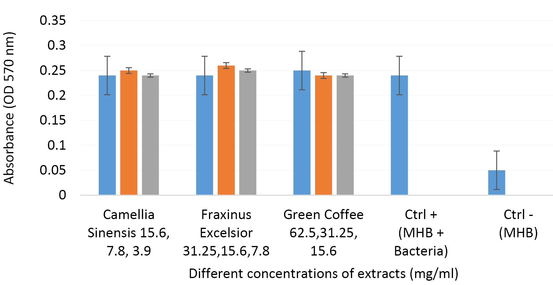
Figure 1.
The OD Values of Different Concentrations of Extracts, Ctrl + (MHB + Bacteria), and Ctrl - (MHB) at 570 nm. Note. OD: Optical density; Ctrl -: Negative control; Ctrl +: Positive control; MHB: Muller Hinton Broth
.
The OD Values of Different Concentrations of Extracts, Ctrl + (MHB + Bacteria), and Ctrl - (MHB) at 570 nm. Note. OD: Optical density; Ctrl -: Negative control; Ctrl +: Positive control; MHB: Muller Hinton Broth
Statistical Analyses
Disk Diffusion and Well Diffusion Methods
Friedman and Wilcoxon tests were performed to evaluate the significance of differences in inhibition zones at various concentrations of extracts. The results indicated a statistically significant variation for C. sinensis extracts across the tested concentrations (P < 0.05).
The Wilcoxon signed-rank test was performed to compare the antibacterial activity between aqueous and ethanolic extracts of C. sinensis at each concentration level. Based on the results, no statistically significant differences were found between the two extract types at all tested concentrations (Z = –1.604 to –1.732, P > 0.05). Therefore, although some numerical differences were observed, they were not statistically meaningful under test conditions.
Comparisons of Minimum Inhibitory and Minimum Bactericidal Concentrations
The Kruskal-Wallis test was applied to compare MIC and MBC values across the different plant extracts. According to the results, there were no statistically significant differences in MIC (Kruskal-Wallis statistic = 8.17, P = 0.147) or MBC (Kruskal-Wallis statistic = 8.17, P = 0.147) across different extracts, indicating that the MIC and MBC values among various plant extracts do not differ significantly.
The Mann-Whitney U test was utilized to compare MIC and MBC values between aqueous and ethanolic extracts across all plant samples. The analysis showed that the U-value was 6.0 for both MIC and MBC comparisons, with a corresponding P value of 1.0 in each case. These results represented no statistically significant difference in antibacterial activity between the two solvent types. Therefore, both types of extracts exhibit similar efficacy in inhibiting the growth of the bacteria.
Discussion
In this study, the antimicrobial and antibiofilm properties of extracts from three medicinal plants were assessed against P. aeruginosa. The extraction was performed using two commonly accepted solvents) water and ethanol), both of which are considered safe for human use. Ethanol, in particular, is widely employed in phytopharmaceutical preparations due to its effectiveness in extracting bioactive compounds (18). The results demonstrated variable antibacterial performance among the tested plant species. While the spot assay (conducted with a low extract concentration of 5 mg/mL) did not reveal any inhibitory effect for any of the extracts, this was expected given the limited dosage applied.
Conversely, in both disc and well diffusion assays, C. sinensis extracts, regardless of solvent type, exhibited notable antibacterial activity against both the reference and drug-resistant P. aeruginosa strains. These findings conform to those of earlier studies that have consistently highlighted the strong antimicrobial potential of polyphenolic compounds present in C. sinensis (19,20). Several studies have reported that catechins, particularly epigallocatechin gallate (identified as a major component in C. sinensis), especially epigallocatechin gallate and epigallocatechin, are mainly responsible for inhibiting bacterial growth (6,7). While prior work has emphasized activity against Gram-positive organisms, our findings highlight the significant efficacy of these extracts even against XDR P. aeruginosa, a Gram-negative, biofilm-forming pathogen. This expands the potential relevance of green tea extracts in multidrug-resistant infections.
In the well diffusion method, the ethanolic extract of F. excelsior could inhibit both tested bacterial strains (with an inhibition zone diameter of 9–11 mm). Conversely, this extract was not affected by the disk diffusion method. This discrepancy may reflect differences in diffusion dynamics between methods or a concentration-dependent threshold of activity. According to some studies, the extracts of F. excelsior have shown antimicrobial activity against bacteria (10,11,21,22). In the study by Amamra et al (22), two extracts from F. excelsior (methanol and petroleum ether extracts) were tested against Bacillus sp. and Pseudomonas sp., and the petroleum ether extract slightly inhibited both strains with inhibition zones of 9.11 ± 0.64 mm and 10.33 ± 0.93 mm, respectively. The methanol extract exhibited good action against Pseudomonas with an inhibition zone of 18.44 ± 1.69 mm, and our findings are partially in line with those reports, though they also underscore the influence of solvent type on extraction efficiency and antimicrobial spectrum. Some chemical compounds isolated from this plant are coumarins, secoiridoids, flavonoids, lignans, phenolic acids, sterols, and triterpenes. These compounds may have different biological effects, such as antioxidant, anti-inflammatory, immunomodulatory, and antimicrobial effects (11).
Green coffee extracts revealed the weakest antibacterial effects in all assays, despite some literature reporting strong activity from roasted or fermented coffee products (23-25). Our findings are consistent with prior reports, noting that green coffee, unlike its roasted counterpart, lacks significant antimicrobial potency, and roasted coffee possesses broad-spectrum antibacterial activity, particularly against Staphylococcus aureus and Streptococcus mutans. The primary contributors to this activity were not chlorogenic acids or caffeine (which have limited antimicrobial power), but rather α-dicarbonyl compounds, such as glyoxal, methylglyoxal, and diacetyl. Caffeine appears to play a supporting role by enhancing the antibacterial effect of these compounds through synergism (23). This contrast reinforces the notion that processing methods significantly impact phytochemical profiles and biological activity. Further supporting this view. Díaz-Hernández et al demonstrated that the ethanolic extracts of roasted coffee and coffee waste were rich in total phenols and exhibited strong antimicrobial properties against clinical isolates (24). Notably, the high MIC/MBC values observed for green coffee extract suggest limited clinical relevance in its crude form, and its use would likely require purification or formulation enhancement.
With regard to antibiofilm activity, the tested extracts did not show a statistically significant reduction in biofilm biomass in P. aeruginosa ATCC 27853 under the intended conditions. However, this observation should be interpreted with caution. The sole use of crystal violet staining measures total biomass (including dead cells and extracellular matrix) without assessing cell viability or metabolic activity. Therefore, it is premature to conclude that the extracts definitively lack antibiofilm properties. Previous studies, especially those utilizing nanoparticles or purified compounds, have reported notable antibiofilm effects (26-28). For example, Ali et al found that C. sinensis silver nanoparticles effectively inhibited biofilms in Candida species, emphasizing the importance of delivery format and concentration (26). In our study, several factors may have contributed to the absence of significant antibiofilm activity, despite the observed antibacterial effects. A primary possibility is that the tested extracts did not interfere with quorum-sensing systems, such as the las and rhl circuits, which are critical in regulating biofilm formation in P. aeruginosa. Additionally, the compounds may not suppress efflux pumps that play a central role in both antibiotic resistance and biofilm persistence. The failure to disrupt bacterial membrane integrity or to penetrate the extracellular polymeric matrix could also have limited the biofilm-inhibitory potential of the extracts. Moreover, considering that only sub-MIC concentrations were tested, it is possible that these levels were insufficient to affect biofilm-specific pathways or structural components. The lack of mechanistic assays (e.g., quantitative polymerase chain reaction analysis of quorum sensing genes [lasI and lasR], efflux pump activity profiling, and membrane permeabilization tests) further restricts our ability to determine the precise reasons for the observed outcome. Future studies should include these targeted evaluations alongside viability-based methods (e.g., resazurin reduction) and imaging tools (e.g., confocal laser scanning microscopy or scanning electron microscopy) to provide a more comprehensive and mechanistic understanding of the antibiofilm potential of these plant-derived extracts.
Based on our findings, C. sinensis extracts demonstrated significant antibacterial activity against both XDR P. aeruginosa and the ATCC 27853 strain, highlighting their potential as alternative antimicrobial agents. Contrarily, the antibacterial activity of F. excelsior was modest, with inhibition zones ranging from 9 mm to 11 mm (below the clinical breakpoints defined by CLSI guidelines), and thus should be interpreted as limited in vitro activity rather than therapeutic efficacy. Nonetheless, plant-derived extracts may offer several advantages, including a lower risk of resistance development due to their multi-target mechanisms and possible use as adjunctive or topical agents, particularly in localized infections. The other advantages are relatively safe solvent systems (water and ethanol) and the potential for further development following compound purification and mechanistic studies.
Nevertheless, additional investigations are required to refine extraction protocols, elucidate the underlying mechanisms of action, and evaluate the clinical safety of these extracts for potential therapeutic application in humans. In addition, other studies should investigate the potential synergistic effects of combining plant extracts with conventional antibiotics to explore their adjuvant therapeutic potential and use high-performance liquid chromatography or liquid chromatography-mass spectrometry to characterize the phytochemical profiles of the extracts. Another point is that the relatively high MIC values observed in this study, particularly for the green coffee extract, may limit its direct therapeutic application in crude form. Future work should focus on purification or advanced formulations to enhance bioavailability and clinical potential.
Limitations of the Study
This study had several limitations. Antibiofilm activity was tested only against P. aeruginosa ATCC 27853, and antibacterial testing relied on a single XDR strain, limiting generalizability. This small sample size reduced statistical power and limited confidence in the observed trends. Additionally, P-values were reported without effect sizes, further limiting interpretability. All assays were in vitro and may not reflect clinical conditions. Biofilm assays were performed in triplicate (n = 3), although using ≥ 6 replicates is recommended for greater reliability. MIC/MBC testing lacked standard antibiotic controls like imipenem, and effect sizes were not reported. Varying extract concentrations across assays hindered direct comparisons, and ethanol-only controls were missing in antibiofilm experiments. No phytochemical profiling or mechanistic studies (e.g., quorum sensing and efflux inhibition) were performed, nor were potential synergistic effects with antibiotics explored.
Conclusion
Overall, our findings demonstrated that C. sinensis and, to a lesser extent, F. excelsior extracts exhibit noteworthy in vitro antibacterial activity against both XDR P. aeruginosa and the ATCC 27853 strain, supporting their potential as sources for alternative antimicrobial agents. However, the relatively high MIC/MBC values for green coffee extracts suggested limited practical applicability in crude form, and the modest inhibition zones for F. excelsior did not meet CLSI breakpoints, warranting a cautious interpretation. It was found that the lack of biofilm inhibition does not preclude potential activity at higher concentrations or via alternative mechanisms but underscores the necessity of viability-based assays in future work. Future research should focus on compound profiling (e.g., total phenolic content, total flavonoid content, or liquid chromatography-mass spectrometry), explore synergistic effects with conventional antibiotics, and use more advanced methods (including gene expression analysis and viability imaging) to validate and expand on these preliminary findings.
Competing Interests
The authors declare that they have no conflict of interests.
Ethical Approval
The Ethics Committees of Dezful University of Medical Sciences, Dezful, Iran (IR.DUMS.REC.1400.055) confirmed this research.
Funding
This research was financially supported by the Student Research Committee of Dezful University of Medical Sciences (1400-SRC-400107). In addition, this article received funding from the Vice-Chancellor of Research of Dezful University of Medical Sciences.
References
- Yousaf H, Mehmood A, Ahmad KS, Raffi M. Green synthesis of silver nanoparticles and their applications as an alternative antibacterial and antioxidant agents. Mater Sci Eng C Mater Biol Appl 2020; 112:110901. doi: 10.1016/j.msec.2020.110901 [Crossref] [ Google Scholar]
- Wang L, Zhong W, Liu B, Pranantyo D, Chan-Park MB. Cationic carbon monoxide-releasing polymers as antimicrobial and antibiofilm agents by the synergetic activity. ACS Appl Mater Interfaces 2023; 15(35):41772-82. doi: 10.1021/acsami.3c02898 [Crossref] [ Google Scholar]
- Xu Q, Chen S, Jiang L, Xia C, Zeng L, Cai X. Sonocatalytic hydrogen/hole-combined therapy for anti-biofilm and infected diabetic wound healing. Natl Sci Rev 2023; 10(5):nwad063. doi: 10.1093/nsr/nwad063 [Crossref] [ Google Scholar]
- Komes D, Horžić D, Belščak A, Ganić KK, Vulić I. Green tea preparation and its influence on the content of bioactive compounds. Food Res Int 2010; 43(1):167-76. doi: 10.1016/j.foodres.2009.09.022 [Crossref] [ Google Scholar]
- Díaz-Puertas R, Álvarez-Martínez FJ, Falco A, Barrajón-Catalán E, Mallavia R. Phytochemical-based nanomaterials against antibiotic-resistant bacteria: an updated review. Polymers (Basel) 2023; 15(6):1392. doi: 10.3390/polym15061392 [Crossref] [ Google Scholar]
- Shedafa RJ, Sempombe J, Nondo RS, Kaale E, Temu M, Imming P. Determination of epigallocatechin gallate (EGCG) and antibacterial activities of commercially available Tanzanian green tea (Camellia sinensis). Tanzan J Sci 2023; 49(2):413-21. doi: 10.4314/tjs.v49i2.12 [Crossref] [ Google Scholar]
- Zihadi MA, Rahman M, Talukder S, Hasan MM, Nahar S, Sikder MH. Antibacterial efficacy of ethanolic extract of Camellia sinensis and Azadirachta indica leaves on methicillin-resistant Staphylococcus aureus and shiga-toxigenic Escherichia coli. J Adv Vet Anim Res 2019; 6(2):247-52. doi: 10.5455/javar.2019.f340 [Crossref] [ Google Scholar]
- Yadav M, Kaushik M, Roshni R, Reddy P, Mehra N, Jain V. Effect of green coffee bean extract on Streptococcus mutans count: a randomised control trial. J Clin Diagn Res 2017; 11(5):ZC68-71. doi: 10.7860/jcdr/2017/25743.9898 [Crossref] [ Google Scholar]
- Tasew T, Mekonnen Y, Gelana T, Redi-Abshiro M, Chandravanshi BS, Ele E. In vitro antibacterial and antioxidant activities of roasted and green coffee beans originating from different regions of Ethiopia. Int J Food Sci 2020; 2020:8490492. doi: 10.1155/2020/8490492 [Crossref] [ Google Scholar]
- Middleton P, Stewart F, Al Qahtani S, Egan P, Orourke C, Abdulrahman A. Antioxidant, antibacterial activities and general toxicity of Alnus glutinosa, Fraxinus excelsior and Papaver rhoeas. Iran J Pharm Res 2005; 4(2):81-6. [ Google Scholar]
- Mahboubi M, Mahdizadeh E, Heidary Tabar R. Chemical composition and antimicrobial activity of Fraxinus excelsior L seeds essential oil. Jundishapur J Nat Pharm Prod 2019; 14(1):e61105. doi: 10.5812/jjnpp.61105 [Crossref] [ Google Scholar]
- Generalić Mekinić I, Skroza D, Ljubenkov I, Katalinić V, Šimat V. Antioxidant and antimicrobial potential of phenolic metabolites from traditionally used Mediterranean herbs and spices. Foods 2019; 8(11):579. doi: 10.3390/foods8110579 [Crossref] [ Google Scholar]
- Sasidharan S, Chen Y, Saravanan D, Sundram KM, Yoga Latha L. Extraction, isolation and characterization of bioactive compounds from plants’ extracts. Afr J Tradit Complement Altern Med 2011; 8(1):1-10. [ Google Scholar]
- Clinical and Laboratory Standards Institute (CLSI). Performance Standards for Antimicrobial Susceptibility Testing: 33th Informational Supplement. CLSI Document M100-S20. Wayne, PA: CLSI; 2023.
- Rajeswaran SA, Arivarasu L, Rajeshkumar S, Thangavelu L. Antimicrobial activity of green tea and mint extract against wound pathogens. J Complement Med Res 2021; 12(3):68-73. doi: 10.5455/jcmr.2021.12.03.09 [Crossref] [ Google Scholar]
- Clinical and Laboratory Standards Institute (CLSI). M07: Methods for Dilution Antimicrobial Susceptibility Tests for Bacteria That Grow Aerobically. 11th ed. Wayne, PA: CLSI; 2018.
- Kırmusaoğlu S. The methods for detection of biofilm and screening antibiofilm activity of agents. In: Kırmusaoğlu S, ed. Antimicrobials, Antibiotic Resistance, Antibiofilm Strategies and Activity Methods. IntechOpen; 2019. doi: 10.5772/intechopen.84411.
- Ganora L. Herbal Constituents: Foundations of Phytochemistry: A Holistic Approach for Students and Practitioners of Botanical Medicine. Herbalchem Press; 2008.
- Dubey N, Mehta A. In vitro study of the antimicrobial property of green tea extract against standard (ATCC) bacterial strains and clinical isolates of methicillin resistant Staphylococcus aureus & multidrug resistant Pseudomonas aeruginosa. Indian J Microbiol Res 2016; 3(3):230-5. doi: 10.5958/2394-5478.2016.00051.0 [Crossref] [ Google Scholar]
- Jaafar ZS. The antimicrobial effects of green tea and lemon juice on Escherichia coli isolated from patients with urinary tract infection in holy Karbala city. Int J Innov Appl Stud 2016; 18(1):318-30. [ Google Scholar]
- Amamra S, Charef NA, Arrar L, Belhaddad O, Khennouf S, Zaim K. Phenolic content, antioxidant and antibacterial effects of fruit extracts of Algerian ash, Fraxinus excelsior. Indian J Pharm Sci 2018; 80(6):1021-8. [ Google Scholar]
- Amamra S, Arrar L, Belhaddad O, Mezaache-Aichour S, Charef N, Haichour N, et al. Antibacterial activity of two extracts of Fraxinus excelsior. 5th Conférence Internationale sur les Méthodes Alternatives de Protection des Plantes. Lille, France: Association Française de Protection des Plantes (AFPP); 2015.
- Daglia M, Papetti A, Grisoli P, Aceti C, Spini V, Dacarro C. Isolation, identification, and quantification of roasted coffee antibacterial compounds. J Agric Food Chem 2007; 55(25):10208-13. doi: 10.1021/jf0722607 [Crossref] [ Google Scholar]
- Díaz-Hernández GC, Alvarez-Fitz P, Maldonado-Astudillo YI, Jiménez-Hernández J, Parra-Rojas I, Flores-Alfaro E. Antibacterial, antiradical and antiproliferative potential of green, roasted, and spent coffee extracts. Appl Sci 2022; 12(4):1938. doi: 10.3390/app12041938 [Crossref] [ Google Scholar]
- Dondapati VB, Bandaru S, Babu SK, Bandaru NR, Perala BK, Voleti A. Antibacterial activity of coffee extract against common human bacterial pathogens in a teaching hospital of semi urban setup. Natl J Physiol Pharm Pharmacol 2023; 13(4):773-7. doi: 10.5455/njppp.2023.13.08381202208092022 [Crossref] [ Google Scholar]
- Ali SG, Jalal M, Ahmad H, Sharma D, Ahmad A, Umar K. Green synthesis of silver nanoparticles from Camellia sinensis and its antimicrobial and antibiofilm effect against clinical isolates. Materials (Basel) 2022; 15(19):6978. doi: 10.3390/ma15196978 [Crossref] [ Google Scholar]
- Jamalifar H, Samadi N, Nowroozi J, Dezfulian M, Fazeli MR. Down-regulatory effects of green coffee extract on las I and las R virulence-associated genes in Pseudomonas aeruginosa. Daru 2019; 27(1):35-42. doi: 10.1007/s40199-018-0234-0 [Crossref] [ Google Scholar]
- José da Costa G, dos Santos RM, Cerávolo IP, Freire GP, Dias Souza MV. Green tea (Camellia sinensis) extract is effective against biofilms of Staphylococcus aureus and Pseudomonas aeruginosa, and interferes on the activity of antimicrobial drugs. Curr Funct Foods 2023; 1(2):E190423216032. doi: 10.2174/2666862901666230419092405 [Crossref] [ Google Scholar]