Avicenna Journal of Clinical Microbiology and Infection. 12(2):81-85.
doi: 10.34172/ajcmi.3614
Original Article
A Cross-Sectional Study of Molecular Identification of Human Papillomavirus in Healthy Women With Normal Cytology in Ahvaz, Iran
Raziyeh Kashisaz Conceptualization, Data curation, Formal analysis, Methodology, Resources, Visualization, Writing – original draft, 1 
Mohammad Roayaei Ardakani Conceptualization, Data curation, Methodology, Project administration, Resources, Supervision, Validation, Visualization, Writing – original draft, Writing – review & editing, 1, *
Seyedeh Elham Rezatofighi Conceptualization, Data curation, Methodology, Resources, Validation, Visualization, Writing – original draft, Writing – review & editing, 1
Manoochehr Makvandi Conceptualization, Data curation, Methodology, Resources, Validation, Visualization, Writing – original draft, Writing – review & editing, 2
Author information:
1Department of Biology, Faculty of Science, Shahid Chamran University of Ahvaz, Ahvaz, Iran
2Department of Virology, Ahvaz Jundishapur University of Medical Science, Ahvaz, Iran
Abstract
Background: Human papillomavirus (HPV) is one of the most common sexually transmitted infections among women. This virus, which has a global distribution, is one of the most serious factors related to cervical cancer (CC). Despite the importance of the virus, there is not enough information about its prevalence in Iran. The aim of this study was to investigate the frequency of HPV and its common types in healthy women referred to HPV screening centers by polymerase chain reaction (PCR) and compare it with cytology results.
Methods: In our cross-sectional study, 271 vaginal and cervical swab samples collected from December 2016 to November 2017 and examined in terms of cytology were received from the laboratory. The samples were related to married women aged 16–72 years. The cytological examination was performed on each specimen. Then, the presence of HPV was investigated using PCR. Positive samples were typed by specific primers of genotypes 6, 11, 16, and 18.
Results: Out of 271 samples, 50 (18.45%) were positive for HPV by PCR. These positive samples demonstrated normal or inflammatory cytology. Among all samples, 1 (0.3%) and 4 (1.47%) cases were positive for HPV-18 and HPV-6, respectively; however, types 11 and 16 were not detected.
Conclusion: Although all samples were negative on the Pap smear, DNA HPV was found in at least 18.45% of the samples. Considering the identification of at least one case of high-risk genotype 18, it is recommended that PCR be used alongside Pap-smear screening, as it enables earlier detection of HPV infections compared to cytology alone, potentially enhancing CC prevention strategies.
Keywords: Human papillomavirus, Pap smear, Cervical cancer, HPV-18, HPV-6, PCR
Copyright and License Information
© 2025 The Author(s); Published by Hamadan University of Medical Sciences.
This is an open-access article distributed under the terms of the Creative Commons Attribution License (
https://creativecommons.org/licenses/by/4.0), which permits unrestricted use, distribution, and reproduction in any medium, provided the original work is properly cited.
Please cite this article as follows: Kashisaz R, Roayaei Ardakani M, Rezatofighi SE, Makvandi M. A cross-sectional study of molecular identification of human papillomavirus in healthy women with normal cytology in Ahvaz, Iran. Avicenna J Clin Microbiol Infect. 2025;12(2):81-85. doi:10.34172/ajcmi.3614
Introduction
Cervical cancer (CC) is the second most common cancer in women. Studies have shown that more than 90% of women who died from this cancer lived in low- and middle-income countries in 2015. The mortality rate due to CC in these countries was eighteen times higher than the rate reported in developed countries (1,2). Human papillomavirus (HPV) has been identified as the main cause of CC, so that viral DNA has been detected in approximately 90% of cervical cancerous lesions. HPV belongs to the Papillomaviridae family (3,4), which has a non-enveloped icosahedral capsid with a circular, double-stranded DNA genome (5). Over 100 types of HPV have been introduced, of which at least 14 are known as high-risk types. Approximately half of these viruses are transmitted through sexual contact and can affect the genitals, mouth, or throat, leading to warts or cancer (6,7). HPV includes high-risk and low-risk groups. Approximately 70% of CC lesions are due to HPV-16 and HPV-18 (high-risk types), while HPV-6 and HPV-11 are the most common agents causing genital warts and are known as low-risk types (8). HPV infection causes cellular proliferation in the epithelium. Infected cells display a wide range of changes, including benign hyperplasia, dysplasia, and invasive carcinoma (9,10). Cervical dysplasia, or cervical intraepithelial neoplasia, is the abnormal growth of cervical surface cells, which can potentially become CC. The cause of intraepithelial neoplasia is the chronic infection of the cervix with HPV, especially infection with high-risk types of 16 or 18 (9). Despite advances in diagnostic and screening methods, as well as vaccine development to prevent and introduce new treatments, this cancer remains an international problem. There is insufficient information related to the prevalence of HPV, the main cause of CC, in Iran; therefore, the present study was conducted to identify the frequency of this virus and its common types in married, healthy women in this region, along with comparing the results of PCR and Pap smear tests.
Materials and Methods
Sample Collection
In this cross-sectional study, the sample size was estimated by a single population proportion formula. Confidence interval, prevalence, and margin of error were considered 95%, 13% (11), and 5%, respectively. The minimum required sample size was 174; nonetheless, 273 samples were investigated to increase the accuracy of the study. To minimize bias, the sampling was performed randomly. The vaginal and cervical swab samples examined in terms of cytology were received from the laboratory. The study stages are shown in Figure 1.
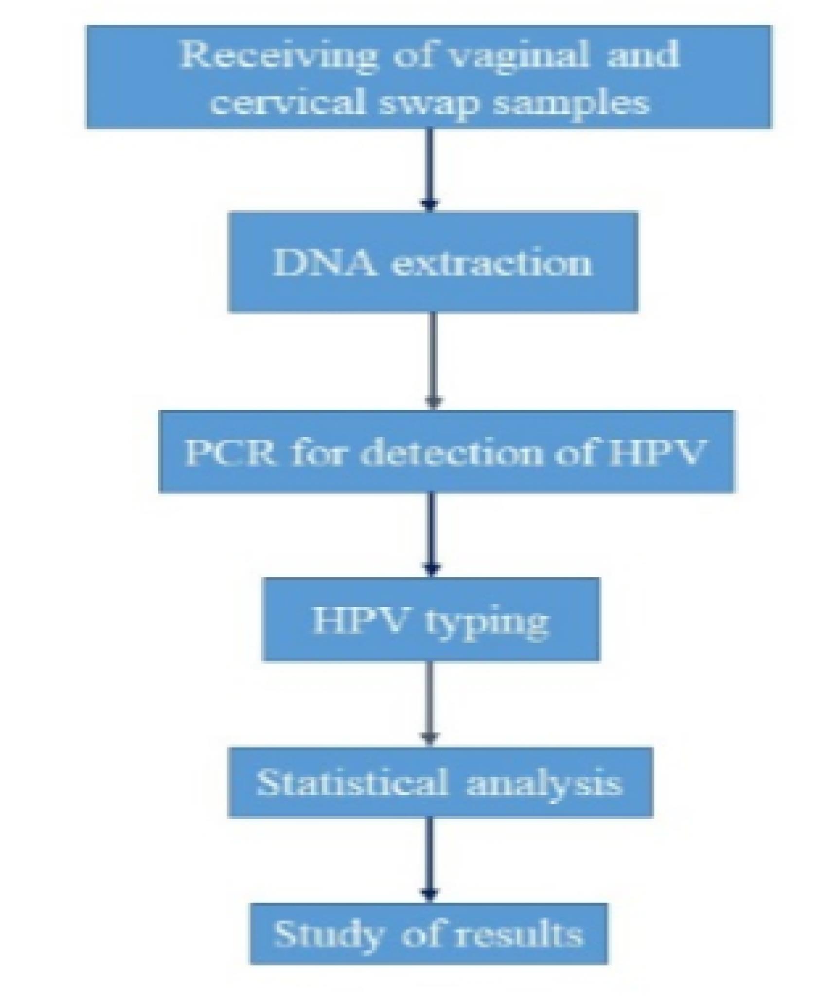
Figure 1.
Flow diagram of study steps
.
Flow diagram of study steps
The cytological examination was performed on each individual specimen. The samples were collected from healthy married women aged 16–72 years old from December 2016 to November 2017. The exclusion criteria included samples related to persons with genital warts, suspected CC, and ulcers in the cervix. The samples were collected with a cytobrush from an endocervix by a gynecologist and were placed in cold phosphate-buffered saline. The results of the cytological examination and demographic characteristics, including age, marital status, number of children, and history of abortion, were received from the laboratory.
Deoxyribonucleic Acid Extraction and Polymerase Chain Reaction
A PCR assay was used to identify the genes L1, E6, and E7 of HPV in vaginal and cervical swab samples. To extract DNA, all samples were vortexed for 20 seconds and centrifuged at 3000 rpm for 10 minutes, and then the supernatant was discarded. The cell pellet was used for subsequent DNA extraction by a kit (CinnaGen, Iran) according to the instructions of the manufacturer. To check the quality of extracted DNA, human β-globin, a housekeeping gene, was amplified using the PCO3 and PCO4 primers (Table 1). The L1 region of the HPV genome was applied to identify HPV-positive samples according to the method of Siddiqa et al (12). HeLa cells infected with HPV-18 were considered as the positive control. The PCR products were electrophoresed on 2.5% agarose gel.
Table 1.
Primers Used for the Detection of β-Globin and the HPV Genome in This Study
|
Primer Name
|
Sequence (5′-3′)
|
Gene
|
Size (bp)
|
Reference
|
PCO3
PCO4 |
ACACAACTGTGTTCACTAGC
CAACTTCATCCACGTTCACC |
β -globin
|
110 |
(12) |
Gp5+
Gp6+ |
TTTGTTACTGTGGTAGATACTACGAA
AAATAAACTGTAAATCATATTC |
L
1
|
150 |
(12) |
| TS16 |
GGTCGGTGGACCGGTCGATG
GCAATGTAGGTGTATCTCCA |
E
6
HPV16
|
96 |
(13) |
| TS18 |
CCTTGGACGTAAATTTTTGG
CACGCACACGCTTGGCAGGT |
L
1
HPV18
|
115 |
(13) |
HF
HR |
TACACTGCTGGACAACATGC
GTGCGCAGATGGGACACAC |
E
6,
E
7
HPV6,11
|
302 |
(11) |
Note. HPV: Human papillomavirus.
Typing of Human Papillomavirus Isolates
The type-specific primers of 6, 11, 16, and 18 were used to determine the genotype of strains. HPV-positive samples were reassigned to PCR by the methods of Ghaffari et al and Gul et al (11,13). The sequence of primers is provided in Table 1. The PCR products were sent to Bioneer Company (South Korea) for sequencing.
Statistical Analysis
The data were analyzed using IBM SPSS statistics software (version 22) using chi-square or Fisher’s exact test. A P value of < 0.05 with a 95% confidence interval was considered statistically significant.
Results
Frequency of Human Papillomavirus in Demographic Groups
A total of 271 vaginal and cervical swab samples were gathered and tested for the presence of HPV infection. The mean age of the subjects was 33 ± 10.08 years. Out of 271 samples, 50 (18.45%) cases were positive for DNA HPV, while they were negative by Pap-smear test and showed normal cytology or inflammation. The mean age of positive HPV cases was 33 ± 10 years. HPV was detected in 17.74% and 18.66% of women with and without abortion, respectively (P> 0.05). The frequency of HPV in women without children or with one child was 48%, and 47.5% of women with more than one child were infected with the virus. In our study, the frequency of HPV was not related to age, abortion, and the number of children (P> 0.05, Table 2).
Table 2.
Frequency of HPV Infection Based on Age, Number of Children, and Abortion
|
Age Groups (N)
|
HPV Positive
N (%)
|
HPV Negative
N (%)
|
P
value (OR)*
|
| 16-25 (48) |
10 (20.83) |
38 (79.17) |
0.992 (0.96-1.02) |
| 26-35 (135) |
25 (18.5) |
110 (81.5) |
| 36-45 (50) |
8 (16) |
42 (84) |
| 46-55 (27) |
6 (22.2) |
21 (77.8) |
| 56-72 (11) |
1 (9.1) |
10 (90.9) |
| Abortion (62) |
11 (17.74) |
51 (82.26) |
0.833 (0.39-1.78) |
| No abortion (209) |
39 (18.66) |
170 (81.34) |
| No or one child (50) |
24 (48) |
26 (52) |
1.108 (0.76-1.60) |
| More than two children (221) |
105 (47.5) |
116 (52.5) |
Note. HPV: Human papillomavirus; OR: Odds ratio.
Frequency of Human Papillomavirus Types
The presence of high-risk (16 and 18) and low-risk (6 and 11) HPV types was investigated in the present study. These types were selected because they are dominant in Iran. Among 271 collected samples, 1 (0.3%) and 4 (1.47%) cases were positive for HPV-18 and HPV-6, respectively; however, types 11 and 16 were not detected in the intended samples.
Discussion
Vaccination against HPV and Pap smear screening tests is optional in Iran. Out-of-marriage sexual relationships are not legal in Iran. These factors can affect the prevalence of HPV and the difference in prevalence in other communities. Unfortunately, accurate statistics on the prevalence of HPV types are unavailable in Iran. In the present study, 271 samples were studied using the cytological method and PCR. Out of 271 specimens, 50 (18.45%) were positive by PCR, while they demonstrated normal cytology or inflammation. According to previous studies, the number of abnormal Pap smears is increasing in Iran (14). HPV is the cause of approximately 70%–84% of CCs (14). Different studies (14-17) have reported various prevalence rates of HPV in Iran (6%–7%, 49.5%, 2.47%, and 24%). The prevalence of HPV also varies from country to country, with 24%, 22.49%, 16.1%, 14.2%, 11%, and 4.74 % in South Africa (18), China (19), Latin America (18), Eastern Europe (18), Brazil (20), and Pakistan (21), respectively. Differences in HPV prevalence rate may be due to diagnostic methods, living conditions, cultural habits, differences in the average age of marriage, and vaccination against HPV. The increase in the prevalence of HPV in Iran may be caused by changes in sexual behavior, increased out-of-marriage sexual relationships, and increased use of tobacco among women (15). As reported in other studies, there was no statistically significant relationship between HPV infection and age; however, 50% of the HPV infections were found in subjects aged from 26–35 years. This is the age when women in Iran usually get married and have sexual relationships. In the universal models, women aged < 29 years and > 50 years are at higher risk of getting a positive HPV test. This creates a U-shaped pattern that can be observed in the present study (22,23). The HPV type’s prevalence also varies from country to country. HPV-18 had a 1.67% prevalence in France (23), 6.25% in Pakistan (21), and 5.7% in India (24). HPV-6 was reported, with prevalence rates of 16%, 1.30%, and 25% in Brazil (20), Southwest China (25), and Pakistan (21), respectively. In positive HPV isolates, one (0.3%) was HPV-18, which is classified as a high-risk type, and four (1.47%) were identified as low-risk HPV-6. None of the positive samples belonged to types 11 and 16. Because our results were limited to HPV types 16, 18, 6, and 11, the 45 remaining HPV isolates became unknown and require further investigation. This is one of the limitations of the study. Another limitation is that the study population could not represent the whole community. In the present study, only people who volunteered for health centers were sampled, which may not demonstrate the broader population. People who had no access to health centers for various reasons were not included in this study. In addition, the data were collected between 2016 and 2017. It may not fully represent the current prevalence of HPV, given possible shifts in risk factors over time. The use of conventional PCR with specific primers, rather than more sensitive methods (e.g., real-time PCR or IVD-certified kits), may have reduced the detection rate, potentially leading to an underestimation of HPV-positive cases. All these factors, along with information and selection biases, can affect the results. Nevertheless, these findings provide a valuable historical reference point for future studies aiming to examine trends in HPV prevalence in this region.
The screening of CC in Ahvaz is currently based on the Pap smear test. However, many asymptomatic HPV infections are ignored due to the false-negative results and the lack of a clear screening program. Therefore, PCR accomplished with a Pap smear can be a useful method for HPV screening.
Conclusion
The primary goal of CC screening is to identify precancerous lesions caused by HPV to remove and prevent invasive cancers from developing. Based on the findings of the present study, most people with HPV have no symptoms or health problems. Accordingly, regular checkups are highly important. The Pap smear test did not show abnormal cells on the cervix, while the PCR test revealed the presence of high-risk types of HPV in cervix samples, which can lead to cancer. Therefore, a combination of HPV vaccination and cervical screening tests by Pap smear and PCR can provide the greatest protection against CC.
Acknowledgements
The authors are very thankful to Shahid Chamran University of Ahvaz for the facilities provided to accomplish the present research project. This article has been extracted from an MSc thesis submitted by Raziyeh Kashisaz.
Competing Interests
The author declared no conflict of interests.
Ethical Approval
In the present study, archived samples of laboratories were used, and therefore there was no need to obtain an ethical code.
Funding
This study received no external funding or support for the publication of this study.
References
- Yang BH, Bray FI, Parkin DM, Sellors JW, Zhang ZF. Cervical cancer as a priority for prevention in different world regions: an evaluation using years of life lost. Int J Cancer 2004; 109(3):418-24. doi: 10.1002/ijc.11719 [Crossref] [ Google Scholar]
- Allanson ER, Schmeler KM. Cervical cancer prevention in low- and middle-income countries. Clin Obstet Gynecol 2021; 64(3):501-18. doi: 10.1097/grf.0000000000000629 [Crossref] [ Google Scholar]
- Williams VM, Filippova M, Soto U, Duerksen-Hughes PJ. HPV-DNA integration and carcinogenesis: putative roles for inflammation and oxidative stress. Future Virol 2011; 6(1):45-57. doi: 10.2217/fvl.10.73 [Crossref] [ Google Scholar]
- Moens U. Human polyomaviruses and papillomaviruses. Int J Mol Sci 2018; 19(8):2360. doi: 10.3390/ijms19082360 [Crossref] [ Google Scholar]
- Graham SV. The human papillomavirus replication cycle, and its links to cancer progression: a comprehensive review. Clin Sci (Lond) 2017; 131(17):2201-21. doi: 10.1042/cs20160786 [Crossref] [ Google Scholar]
- Coser J, da Rocha Boeira T, Simon D, Kazantzi Fonseca AS, Ikuta N, Lunge VR. Prevalence and genotypic diversity of cervical human papillomavirus infection among women from an urban center in Brazil. Genet Mol Res 2013; 12(4):4276-85. doi: 10.4238/2013.February.19.3 [Crossref] [ Google Scholar]
- Bahramabadi R, Honarvar Z, Iranpour M, Kazemi Arababadi M, Dehesh T, Dabiri B. Epidemiological study of various HPV strains in cervical fluid samples in south-eastern Iran, 2018-2020. Arch Iran Med 2021; 24(9):678-83. doi: 10.34172/aim.2021.97 [Crossref] [ Google Scholar]
- Mutombo AB, Benoy I, Tozin R, Bogers J, Van Geertruyden JP, Jacquemyn Y. Prevalence and distribution of human papillomavirus genotypes among women in Kinshasa, the Democratic Republic of the Congo. J Glob Oncol 2019; 5:1-9. doi: 10.1200/jgo.19.00110 [Crossref] [ Google Scholar]
- Balasubramaniam SD, Balakrishnan V, Oon CE, Kaur G. Key molecular events in cervical cancer development. Medicina (Kaunas) 2019; 55(7):384. doi: 10.3390/medicina55070384 [Crossref] [ Google Scholar]
- Kadaja M, Silla T, Ustav E, Ustav M. Papillomavirus DNA replication - from initiation to genomic instability. Virology 2009; 384(2):360-8. doi: 10.1016/j.virol.2008.11.032 [Crossref] [ Google Scholar]
- Ghaffari SR, Sabokbar T, Mollahajian H, Dastan J, Ramezanzadeh F, Ensani F. Prevalence of human papillomavirus genotypes in women with normal and abnormal cervical cytology in Iran. Asian Pac J Cancer Prev 2006; 7(4):529-32. [ Google Scholar]
- Siddiqa A, Zainab M, Qadri I, Bhatti MF, Parish JL. Prevalence and genotyping of high-risk human papillomavirus in cervical cancer samples from Punjab, Pakistan. Viruses 2014; 6(7):2762-77. doi: 10.3390/v6072762 [Crossref] [ Google Scholar]
- Gul S, Murad S, Javed A. Prevalence of high-risk human papillomavirus in cervical dysplasia and cancer samples from twin cities in Pakistan. Int J Infect Dis 2015; 34:14-9. doi: 10.1016/j.ijid.2015.02.018 [Crossref] [ Google Scholar]
- Haghshenas M, Golini-Moghaddam T, Rafiei A, Emadeian O, Shykhpour A, Ashrafi GH. Prevalence and type distribution of high-risk human papillomavirus in patients with cervical cancer: a population-based study. Infect Agent Cancer 2013; 8(1):20. doi: 10.1186/1750-9378-8-20 [Crossref] [ Google Scholar]
- Mobini Kesheh M, Keyvani H. The prevalence of HPV genotypes in Iranian population: an update. Iran J Pathol 2019; 14(3):197-205. doi: 10.30699/ijp.2019.90356.1861 [Crossref] [ Google Scholar]
- Noor M, Ghani E, Niazi SK, Khadim MT. Molecular detection of human papillomavirus infection of uterine cervix among women of reproductive age. J Virol Antivir Res 2019; 8(1):188. doi: 10.4172/2324-8955.1000188 [Crossref] [ Google Scholar]
- Farahmand Z, Soleimanjahi H, Garshasbi M, Hasanzadeh M, Zafari E. Distribution of the most common types of HPV in Iranian women with and without cervical cancer. Women Health 2021; 61(1):73-82. doi: 10.1080/03630242.2020.1822490 [Crossref] [ Google Scholar]
- Bruni L, Diaz M, Castellsagué X, Ferrer E, Bosch FX, de Sanjosé S. Cervical human papillomavirus prevalence in 5 continents: meta-analysis of 1 million women with normal cytological findings. J Infect Dis 2010; 202(12):1789-99. doi: 10.1086/657321 [Crossref] [ Google Scholar]
- Zhong TY, Zhou JC, Hu R, Fan XN, Xie XY, Liu ZX. Prevalence of human papillomavirus infection among 71,435 women in Jiangxi province, China. J Infect Public Health 2017; 10(6):783-8. doi: 10.1016/j.jiph.2017.01.011 [Crossref] [ Google Scholar]
- Miranda PM, Pitol BC, Moran MS, Silva NN, Felix PM, Lima-Filho JL. Human papillomavirus infection in Brazilian women with normal cervical cytology. Genet Mol Res 2012; 11(2):1752-61. doi: 10.4238/2012.June.29.8 [Crossref] [ Google Scholar]
- Aziz H, Iqbal H, Mahmood H, Fatima S, Faheem M, Sattar AA. Human papillomavirus infection in females with normal cervical cytology: genotyping and phylogenetic analysis among women in Punjab, Pakistan. Int J Infect Dis 2018; 66:83-9. doi: 10.1016/j.ijid.2017.11.009 [Crossref] [ Google Scholar]
- Liang LA, Tanaka LF, Radde K, Bussas U, Ikenberg H, Heideman DA. Population-based age- and type-specific prevalence of human papillomavirus among non-vaccinated women aged 30 years and above in Germany. BMC Infect Dis 2024; 24(1):1008. doi: 10.1186/s12879-024-09827-7 [Crossref] [ Google Scholar]
- Adenis A, Dufit V, Douine M, Najioullah F, Molinie V, Catherine D. The singular epidemiology of HPV infection among French Guianese women with normal cytology. BMC Public Health 2017; 17(1):279. doi: 10.1186/s12889-017-4181-3 [Crossref] [ Google Scholar]
- Sontakke BR, Ambulkar PS, Talhar S, Shivkumar PV, Bharambe MS, Pal A. Molecular genetic study to detect prevalence of high-risk human papillomavirus strains (type 16 and 18) in cervical lesions and asymptomatic healthy subjects of rural central India. J Cytol 2019; 36(1):32-7. doi: 10.4103/joc.Joc_10_18 [Crossref] [ Google Scholar]
- Hu JP, Wang JL, Li Y, Feng Y, Tian CQ, Zhang GH. Prevalence and genotype distribution of human papillomavirus infection among 66000 women from 2014 to 2023 in the plateau region of Southwest China. Virol J 2024; 21(1):176. doi: 10.1186/s12985-024-02447-2 [Crossref] [ Google Scholar]