Avicenna Journal of Clinical Microbiology and Infection. 12(1):21-28.
doi: 10.34172/ajcmi.3591
Original Article
Investigation of Antibacterial Properties and Healing Effects of Silver Nanoparticles Synthesized from Nettle Extract for Treating Burn Infections in Mice
Nader Kazemi Conceptualization, Data curation, Methodology, 1, * 
Mahdi Arfaei Formal analysis, Writing – original draft, 2
Masoud Kaboutari Supervision, Writing – review & editing, 2
Author information:
1Nanobiotechnology Research Center, Zanjan Branch, Islamic Azad University, Zanjan, Iran
2Department of Biology, Faculty of Basic Sciences, Zanjan Branch, Islamic Azad University, Zanjan, Iran
Abstract
Background: Infections in burns caused by Staphylococcus aureus, Escherichia coli, and Pseudomonas aeruginosa are prevalent and may result in severe health issues. Conventional treatments often struggle with bacterial resistance, highlighting the need for alternative approaches. Due to their strong antimicrobial properties, silver nanoparticles have attracted significant attention. The eco-friendly synthesis of these nanoparticles using plant extracts, such as Urtica dioica, is emerging as a promising strategy. This study aimed to synthesize silver nanoparticles using U. dioica extract through a green method and evaluate their antimicrobial activity and wound healing effects against burn infections.
Methods: Acetonic extracts of U. dioica were mixed with silver nitrate to produce silver nanoparticles. Their synthesis was confirmed using X-ray diffraction (XRD) and dynamic light scattering (DLS). The antimicrobial potential of the nanoparticles and plant extract was evaluated using minimum bactericidal concentration (MBC) and minimum inhibitory concentration (MIC) assays. Rats with burn wounds infected by bacteria were treated with ointments containing either the nanoparticles or the plant extract.
Results: Silver nanoparticles synthesized from U. dioica showed strong antibacterial effects against all three bacterial strains, while the extract alone was effective only against S. aureus and E. coli. The nanoparticles also demonstrated superior wound healing effects compared to the extract.
Conclusion: Silver nanoparticles demonstrated superior antimicrobial and wound healing properties compared to U. dioica extract, making them a strong candidate for burn infection treatment. However, silver nanoparticles are more effective against a broader range of bacteria and demonstrate enhanced wound healing potential, making them strong candidates for treating burn infections.
Keywords: Microdilution method, Minimum bactericidal concentration (MBC), Minimum inhibitory concentration (MIC), Silver nanoparticles, Urtica dioica
Copyright and License Information
© 2025 The Author(s); Published by Hamadan University of Medical Sciences.
This is an open-access article distributed under the terms of the Creative Commons Attribution License (
https://creativecommons.org/licenses/by/4.0), which permits unrestricted use, distribution, and reproduction in any medium, provided the original work is properly cited.
Please cite this article as follows: Kazemi N, Arfaei M, Kaboutari M. Investigation of antibacterial properties and healing effects of silver nanoparticles synthesized from nettle extract for treating burn infections in mice. Avicenna J Clin Microbiol Infect. 2025; 12(1):21-28. doi:10.34172/ajcmi.3591
Introduction
Burns are among the significant causes of skin damage, with approximately 300 000 individuals worldwide losing their lives annually due to severe burn injuries (1). Burns can increase the risk of tissue infection, delay skin healing, and lead to blood infections or sepsis (2). Some bacterial species such as Escherichia coli, Staphylococcus aureus, and Pseudomonadaceae can colonize burn wounds, particularly during extended hospital stays. These bacteria exploit opportunities at wound sites, weakening the immune system and rapidly replicating due to acquired antibiotic resistance (3). S. aureus is a Gram-positive facultative anaerobic bacterium that can sometimes become aerobic (4). This bacterium can be part of the normal flora (5) and is a common agent in hospital-acquired infections, causing burns and surgical infections (6). E. coli is a facultative anaerobic Gram-negative bacterium (7) commonly associated with infections shared between humans and animals. The emergence of antibiotic-resistant strains poses a public health concern (8). P. aeruginosa, a Gram-negative opportunistic bacterium, significantly impacts immunocompromised individuals and is linked to burn infections and cystic fibrosis (9). In burn injuries, damaged skin layers and immune cells create a conducive environment for P. aeruginosa to establish colonies, potentially leading to infection, septic shock, and even death (10). Infection is a significant challenge (11). Various approaches can be used for treating these issues, with silver nanoparticles serving as an antimicrobial agent for burn-related infections and an alternative to antibiotics (12). Due to their high stability, low reactivity, potent antimicrobial properties, and healing effect, silver nanoparticles have been extensively studied (13). Nanomaterial-based drug delivery carriers with antimicrobial behavior provide novel platforms for treating various inflammatory diseases such as burn infections (14). Moreover, plants serve as valuable sources of antimicrobial agents due to their rich composition of bioactive molecules, including amino acids, vitamins, phytochemicals, and polyphenolic compounds such as flavonoids and phenolic acids, which have gained considerable scientific interest. U. dioica, a well-known medicinal herb, contains a variety of essential minerals and antimicrobial constituents like flavonoids and phenolic acids, making it a strong candidate for the development of therapeutic formulations for burn infections (15). Nanotechnology, which focuses on materials structured at the atomic or molecular level, has emerged as a crucial tool in medical sciences, particularly for combating microbial pathogens. Certain naturally occurring antibacterial elements, such as zinc and silver, exhibit significantly improved antimicrobial and wound-healing properties when processed into nanoscale structures (16).
Objectives
This study aimed to compare the antimicrobial and healing properties of silver nanoparticles synthesized from the acetonic extract of U. dioica with those of the extract itself in treating burn infections caused by S. aureus, Escherichia coli, and Pseudomonas aeruginosa in mice. The macrodilution method was employed to determine the minimum inhibitory concentration (MIC) and minimum bactericidal concentration (MBC). Furthermore, the silver nanoparticles were characterized through material analysis techniques.
Materials and Methods
Preparation of Acetonic Extract of Nettle
First, U. dioica was collected and dried. Next, 40 g of ground sample was weighed using a laboratory scale and placed into a 500 mL Erlenmeyer flask. Then, acetone was added at a ratio of 1:10 (mass of powdered plant: acetone volume), and the flask was sealed with parafilm and stored in a refrigerator at 4 °C for 48 hours. After 48 hours, the solutions were filtered through 4 layers of sterile gauze and centrifuged for 4 minutes at 2500 rpm. Finally, the extracts were diluted and poured into Petri dishes to dry at room temperature, away from sunlight (17).
Synthesis of Silver Nanoparticles
Silver nanoparticles were synthesized using a green method. First, a 0.1 M solution of silver nitrate was prepared in a beaker. Then, 4 mL of the filtered extract was added dropwise to the solution using a PTFE filter to purify the extract from suspended particles. After stirring for 1-2 hours at 80 °C, signs of synthesis were observed, the color of the solution changed to black, and the solution status changed from liquid to foam. The pH of the solution was adjusted by adding 5 mL of NaOH. The synthesized solution was dried in an 80 °C oven for 48 hours. Afterwards, the nanoparticle films were stored in a refrigerator for material and antimicrobial analysis (18). It is necessary to mention that alternative methods, such as co-precipitation and chemical methods, also exhibit antimicrobial activity (19).
Material Characterization
Silver nanoparticles (Ag⁺), ranging in size from 20 to 80 nm, were successfully synthesized using the acetonic solution derived from U. dioica. Their structural and optical properties were analyzed through UV-visible spectroscopy, X-ray diffraction (XRD) analysis, and Zeta potential measurements. Following characterization, the antimicrobial and wound-healing capabilities of the synthesized nanoparticles were evaluated (19).
Preparation of Staphylococcus aureus, Escherichia coli, and Pseudomonas aeruginosa Strains
Staphylococcus aureus strain ATCC292113, E. coli strain ATCC25922, and P. aeruginosa strain ATCC1542 were obtained from the Microbial Bank of the Nanobiotechnology Research Center at Islamic Azad University, Zanjan branch. These pathogenic bacteria were sub-cultured on Mueller Hinton Agar medium.
Determination of MBC and MIC
MBC and MIC values were determined for three antimicrobial agents: silver nanoparticles, acetonic extract of U. dioica combined with nanoparticles, and the extract alone. First, stock solutions for each agent were prepared at a concentration of 500 mg/mL by dissolving the extract film in dimethyl sulfoxide (DMSO) (Merck, Germany). After adding Mueller-Hinton broth (Merck) to each well plate (SPL), serial dilutions were prepared, starting with a concentration of 250 mg/mL. The first step involved adding 500 µL of the stock solution to 500 µL of Mueller-Hinton broth, followed by 500 µL of bacterial suspension. The concentration in each subsequent well was halved compared to the previous one, with the dilution carried out using an Eppendorf pipette, transferring 500 µL from one well to the next. Then, the cellular concentrations of bacterial cultures were adjusted to a cell density of 1.5 × 10⁸ cells/mL (0.5 McFarland standard) (Iran Baharafshan). Afterwards, they were added to their respective well plates, which were subsequently incubated (Memmert) at 37 ºC for 24 hours. After incubation, all the well plates were examined using positive and negative controls. Finally, the MBC and MIC values were determined by culturing the bacteria on the plates containing Mueller Hinton agar medium (20).
Animal Model
A total of 45 adult laboratory rats weighing approximately 200 ± 10 g were used in this study. The rats were obtained from the Faculty of Medical Sciences and transferred to the animal laboratory at the Nanobiotechnology Research Center of Islamic Azad University, Zanjan Branch. They were allowed to acclimate for 48 hours under controlled conditions with a 12-hour light/dark cycle, a temperature of 24 ºC-25 ºC, and a humidity of about 51%. The rats were provided with standard food and water. Afterwards, the study included three main categories based on bacterial strains. The first category was S. aureus, which consisted of five subgroups: (1) control group, three mice that received no treatment, (2) acetonic extract group, three mice that were treated with acetonic extract, (3) silver nanoparticles group, three mice that were treated with silver nanoparticles, (4) Eucerin group, three mice that received a moisturizing ointment without antimicrobial properties, and (5) silver nanoparticles + acetonic extract group, three mice that were treated with a combination of silver nanoparticles and acetonic extract. The second category was E. coli, and the third was P. aeruginosa, both of which followed the same subgrouping as the S. aureus category. To induce burns, the rats were anesthetized by the intraperitoneal administration of thiopental (4 mg/kg). Then, the hair on the back of their necks was shaved, and a molten circle-shaped metal with a diameter of 1 cm was placed on the shaved area for 3-4 seconds. After the rats regained consciousness, they were housed in clean and disinfected cages. On the following day, the wounds were inoculated with bacterial suspensions containing 6 × 10⁸ cells/mL. After 24 hours, treatment was initiated using 1 g of Eucerin ointment for 6 days (0.5 g of ointment per day, administered three times). On the 4th day, samples were collected from each wound using the punching method and cultured in blood agar for colony counting (21). This research was conducted in accordance with international standards. The study was also approved by the Ethics Committee of Islamic Azad University, Zanjan branch (IR.IAU.Z.REC.1400.001).
Statistical Analysis
The gathered data were evaluated using a one-way ANOVA and the LSD test. A significance level of P < 0.01 was considered for all comparisons (22).
Results
UV-Visible Spectrophotometry
The UV-Visible spectrophotometry analysis of the acetonic extract of U. dioica containing silver nitrate is shown in Figure 1B. The distinct absorption peak in the spectrum verifies the effective production of silver nanoparticles. Spectrophotometric measurements were conducted after the nitrate-containing extract was kept under laboratory conditions for 24 hours. In contrast, the wavelength pattern of the acetonic extract alone exhibited a downward trend, as illustrated in Figure 1A. These results confirm the successful fabrication of silver nanoparticles utilizing the acetonic extract of U. dioica, as evidenced by the characteristic absorption peak. Additionally, the data in Text Box 1 illustrate the presence of nanoparticles. It is noteworthy that the wavelength associated with the extract fluctuated due to the presence of naturally occurring impurities (Figure 1).
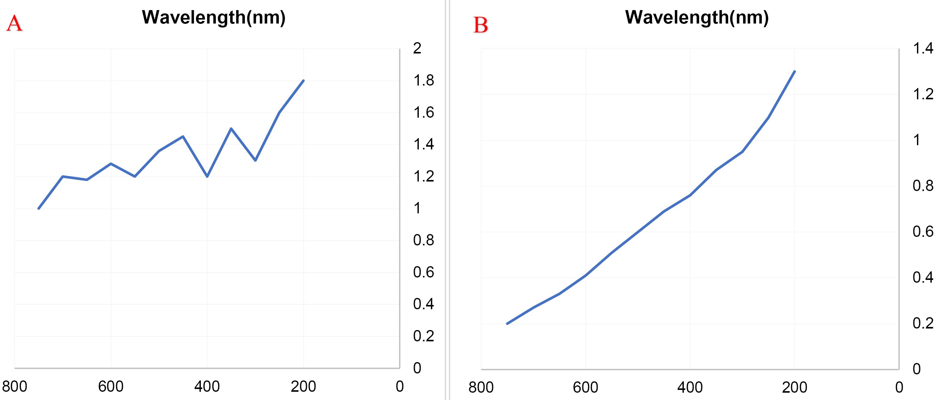
Figure 1.
Average Absorbance of the Acetonic Extract of Urtica dioica Following a 24-hour Period (A), Average Absorbance of Silver Nanoparticles Synthesized from Acetonic Extract of U. dioica after 24 Hours (B). This figure illustrates the mean absorbance of silver nanoparticles synthesized from the acetonic extract, alongside the absorbance spectrum of the extract itself
.
Average Absorbance of the Acetonic Extract of Urtica dioica Following a 24-hour Period (A), Average Absorbance of Silver Nanoparticles Synthesized from Acetonic Extract of U. dioica after 24 Hours (B). This figure illustrates the mean absorbance of silver nanoparticles synthesized from the acetonic extract, alongside the absorbance spectrum of the extract itself
X-Ray Diffraction Analysis
The XRD analysis of silver nanoparticles derived from the acetonic extract of U. dioica revealed corresponding peaks at angles of 29º, 38º, 48º, and 65º (Figure 2). These peaks are consistent with the standard XRD pattern of silver, confirming the successful synthesis of silver nanoparticles. Additionally, the obtained pattern indicates that the synthesized particles possess a specific size distribution, which can be further analyzed for potential applications. This section summarizes the XRD data, emphasizing the key findings that validate the synthesis of silver nanoparticles with the acetonic extract of U. dioica. It is worth mentioning that these peaks were reached by measuring crystallography features and these are the same as those of standard graphs.
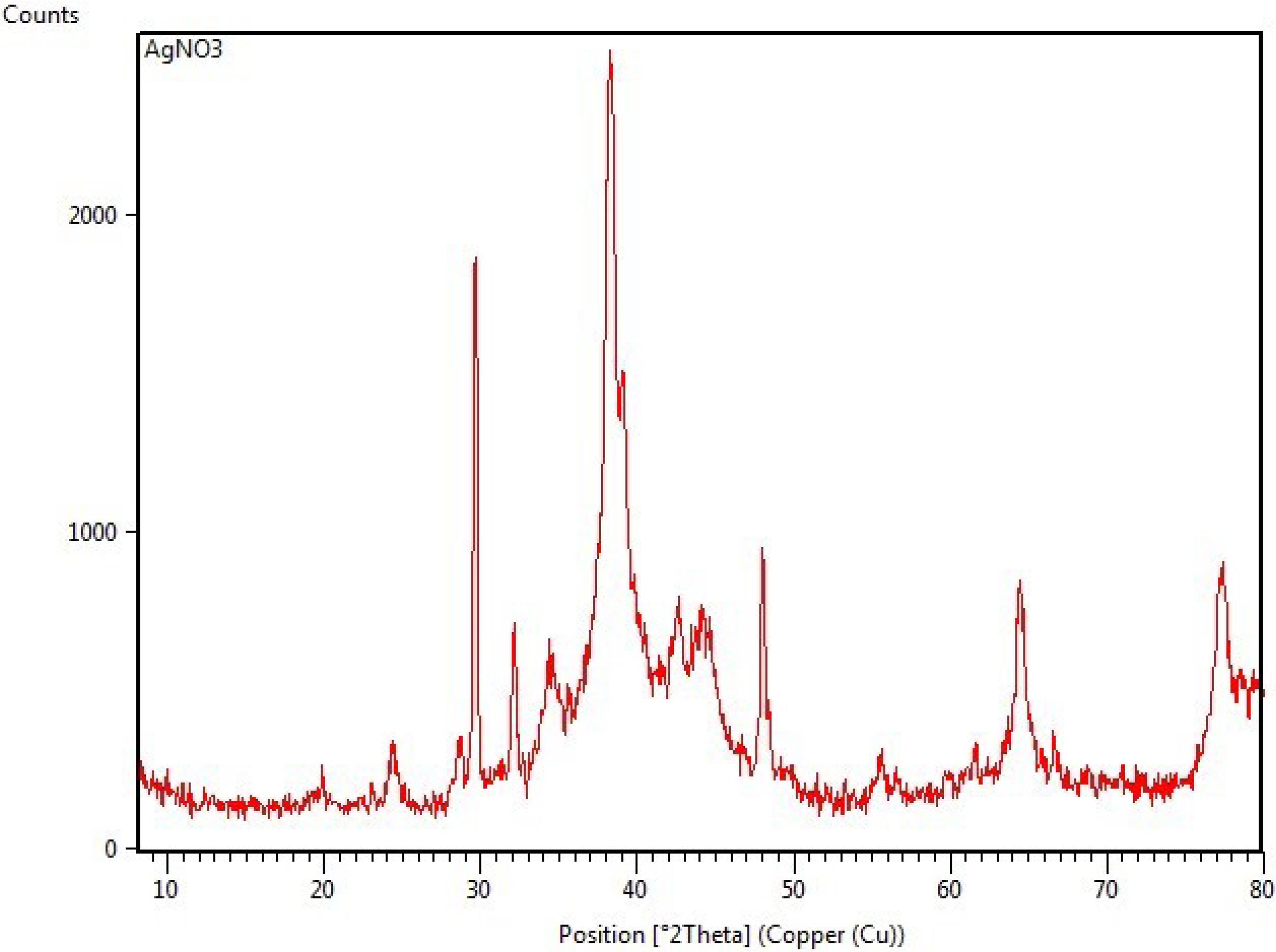
Figure 2.
The XRD figure of silver nano particles there are four main peaks which are same with standard graph of silver nanoparticles. These peaks positions include 29, 38, 48 and 65.
.
The XRD figure of silver nano particles there are four main peaks which are same with standard graph of silver nanoparticles. These peaks positions include 29, 38, 48 and 65.
Zeta Potential of Nanoparticles
The zeta potential data revealed that the synthesized silver nanoparticles exhibited a zeta potential of -50.05 mV at a temperature of 25.1ºC (A) and -47.00 mV at 25.2ºC (B). The dynamic light scattering (DLS) test determines the hardness, amount, and size of the synthesized nanoparticles. Additionally, this test can identify the size of nanoparticles. The quality of the data obtained from this analysis is evaluated by analyzing the autocorrelation function (ACF = g2) and its goodness of fit based on the characteristics of static light scattering and particle size using machine intervals (Frequency Undersize). Key quality indicators in this analysis include: (a) The ACF line and curve must reach 1.0. (b) The ACF sum of squares (SOS) for monodisperse samples must be less than 100 nm. Based on the described criteria, particles with a size of 70–80 nm and an ACF of 1.0 indicate high-quality nanoparticles with proper medical correlation (C). These values indicate low mobility, suggesting that the synthesized particles are stable and resistant to agglomeration. This revised section highlights the key findings from the zeta potential analysis, emphasizing the stability of the synthesized silver nanoparticles. The results demonstrate that nanoparticles exhibit minimal agglomeration, making them suitable for potential applications in various fields.
Determination of MBC and MIC
MBC and MIC values were determined for three antimicrobial agents tested against S. aureus, E. coli, and P. aeruginosa. These agents included silver nanoparticles (AgNPs), the acetonic extract of U. dioica, and AgNPs combined with the acetonic extract of U. dioica.
Silver Nanoparticles
AgNPs were effective at the lowest concentrations against all three bacterial species, including S. aureus, E. coli, and P. aeruginosa.
Silver Nanoparticles Combined with Acetonic Extract of Urtica dioica
The combination of AgNPs and acetonic extract showed effectiveness at minimal concentrations against S. aureus and E. coli, while it exhibited a moderate effect on P. aeruginosa.
Acetonic Extract of Urtica Dioica
The acetonic extract of U. dioica alone demonstrated efficacy at medium concentrations against S. aureus and E. coli, and at higher concentrations against P. aeruginosa.
The results are summarized in Table 1 for S. aureus, E. coli, and P. aeruginosa, respectively. These findings indicate that AgNPs and AgNPs combined with the acetonic extract are more effective against the tested bacterial species compared to the acetonic extract alone.
Table 1.
MBC and MIC of the Antimicrobial Agents against Staphylococcus aureus, Escherichia coli, and Pseudomonas aeruginosa
|
Bacteria
|
Antimicrobial Agents
|
MBC
|
MIC
|
|
Staphylococcus aureus
|
Silver nanoparticles |
3.9 mg/mL |
1.9 mg/mL |
|
Staphylococcus aureus
|
Silver NPs with extract |
3.9 mg/mL |
1.9 mg/mL |
|
Staphylococcus aureus
|
Acetonic extract |
15.6 mg/mL |
7.8 mg/mL |
|
Escherichia coli
|
Silver nanoparticles |
3.9 mg/mL |
1.9 mg/mL |
|
Escherichia coli
|
Silver NPs with extract |
3.9 mg/ mL |
1.9 mg/mL |
|
Escherichia coli
|
Acetonic extract |
15.6 mg/mL |
7.8 mg/mL |
|
Pseudomonas aeruginosa
|
Silver nanoparticles |
3.9 mg/mL |
1.9 mg/mL |
|
Pseudomonas aeruginosa
|
Silver NPs with extract |
15.6 mg/mL |
7.8 mg /mL |
|
Pseudomonas aeruginosa
|
Acetonic extract |
31.2 mg/mL |
15/6 mg/mL |
Animal Model
In the animal model, the results were expressed in colony forming units (CFUs). The groups treated with silver nanoparticles against all bacteria showed a significant difference compared to the control group (P < 0.01). In addition, a significant difference was observed between these groups and those that received both nanoparticles and plant extract, as well as those treated with the acetonic extract alone (P < 0.01), as presented in Table 2.
Table 2.
Effect of the AntimicrobialAgents on the Number of Staphylococcus aureus, Escherichia coli, and Pseudomonas aeruginosa Colonies
|
Group
|
Staphylococcus aureus
Mean±SD (105)
|
Escherichia coli
Mean±SD (105)
|
Pseudomonas aeruginosa
Mean±SD (105)
|
| Treatment with silver nanoparticles |
3.66 × 10⁵ ± 1.15* |
4.66 × 10⁵ ± 0.57* |
7.66 × 10⁵ ± 1.52* |
| Treatment with silver NPs and extract |
5 × 10⁵ ± 2.64* |
5.66 × 10⁵ ± 1.52* |
10.66 × 10⁵ ± 2.08* |
| Treatment with acetonic extract |
8.33 × 10⁵ ± 8.52* |
9.66 × 10⁵ ± 1.52* |
19.33 × 10⁵ ± 1.52* |
| Treatment with Eucerin |
20.33 × 10⁵ ± 2.51* |
21 × 105 ± 2* |
25.66 × 10⁵ ± 2.08* |
| Control and no treatment |
275 × 106 ± 2.65 |
294.33 ± 106 ± 7.37 |
295 × 106 ± 4.35 |
* Indicates a significant difference with the control and no treatment groups (P < 0.01).
The healing process in the extract group was observed to occur within a short time, and a similar effect was seen in the groups treated with silver nanoparticles after 6 days. In contrast, the control groups required 21 days for healing, while the groups treated with a combination of silver nanoparticles and extract took 8 days to heal.
The antimicrobial activity and healing effects in the groups treated with silver nanoparticles were significantly more effective than other treatment groups in this study.
Discussion
Burn wounds are vulnerable to opportunistic colonization by both endogenous and exogenous microorganisms. Factors like the patient’s age, the severity of the burn, and the depth of the wound, combined with microbial variables such as the species and quantity of microorganisms, their enzyme and toxin production, and motility, all affect the risk of infection in burn wounds. These infections can be categorized based on the type of organism, the depth of invasion, and the response of the tissue. Common pathogens responsible for burn wound infections include E. coli, S. aureus, and P. aeruginosa (23,24). Both green and chemical methods are commonly used to synthesize silver nanoparticles; however, the green method is more eco-friendly as it utilizes plant extracts. Ahmad et al studied the green synthesis of silver nanoparticles and their antimicrobial activity. In the present study, the methods of synthesis, characterization of nanoparticles, and antimicrobial activity were aligned with the findings of the study conducted by Ahmad (25). The results from animal studies and the antimicrobial activity of silver nanoparticles against burn infections caused by S. aureus and E. coli were consistent with the study conducted by Wasef et al, which investigated the effects of silver nanoparticles on burn wound healing in a mouse model (26). However, evaluating the cytotoxicity of nanoparticles in medical applications is crucial in assessing their safety (27). One of the limitations of this study is the absence of cytotoxicity assays such as the MTT assay or other related tests. Although our findings, consistent with those of Wasef et al, confirm the antimicrobial properties of the nanoparticles, further investigations are necessary to assess their potential biomedical applications comprehensively. According to the study conducted by Jagtap et al, additional cytotoxicity evaluations are recommended to ensure the safety and efficiency of these nanoparticles in biomedical environments (26,28). The XRD analysis confirmed the crystalline nature of the synthesized nanoparticles, as indicated by the characteristic diffraction peaks. A comparison with previous studies, such as those conducted by Abdulsahib et al and Hassan Afandy et al, demonstrated a strong similarity in peak positions and intensities, suggesting comparable crystallinity. Minor variations observed in the diffraction patterns may stem from differences in synthesis parameters, precursor concentrations, and particle size distributions. These results support the reliability of our XRD findings and further confirm the successful formation of crystalline nanoparticles (19,29). However, the UV-Vis spectrophotometry results in the present study differ from those of previous studies. In the study conducted by Abdulsahib et al, the peak was recorded at 400 nm (19), while in the study conducted by Ansari et al, the absorption spectrum was observed in the range of 420–440 nm (30). Additionally, in the study by Tesfaye et al, the absorption spectrum was found in the range of 411–430 nm (31). In contrast, in the current study, the peak was observed below 400 nm. This effect has been investigated in the studies by Ansari et al and Tesfaye et al, where various factors such as different temperatures, synthesis times, pH, solvent concentration, plant extract volume, and nanoparticle synthesis time were found to influence the absorption spectrum in different ranges (30,31). Zeta potential plays a crucial role in determining the stability of nanoparticles, as it directly affects their tendency to aggregate or remain dispersed (32). In the present study, the synthesized silver nanoparticles exhibited a zeta potential of -50.05 mV (Figure 3A) and + 47 mV (Figure 3B), indicating strong repulsion between particles. These findings are consistent with the results reported by Alzubaidi et al, who recorded zeta potential values of -44.5 and + 231.8 mV (33), and by Netala et al, who reported values of + 44 mV and -19.65 mV (34). The differences in the reported values between this study and previous ones may be due to variations in factors such as nanoparticle size, solvent type, or temperature. The results of this study, along with previous studies, indicate that the synthesized nanoparticles have diverse applications in biomedical fields (33,34). Antimicrobial tests were performed with high sensitivity, in line with the study conducted by Kowalska-Krochmal et al (20) on microdilution and macro-serial dilution. Studies have shown that smaller sample sizes reduce statistical power, making results less reliable (35). Additionally, variations in the pathogen spectrum studied may lead to inconsistencies and hinder the generalization of results in clinical settings (36). The concentration of the bacterial inoculum and the volume of the medium were consistent with the studies by Andrews (37) and Wiegand et al (38). However, inadequate sample size and pathogen spectrum can affect the clinical relevance and reproducibility of findings. Studies have shown that smaller sample sizes reduce statistical power, making results less reliable (35). Additionally, variations in the pathogen spectrum studied may lead to inconsistencies and hinder the generalization of results in clinical settings (36). Therefore, increasing sample sizes and broadening the pathogen spectrum would enhance the reliability and applicability of nanoparticle-based studies. Dağlıoğlu et al studied the biosynthesis of silver nanoparticles derived from Nettle leaf extract, and the materials analysis was consistent with the current study. However, the antimicrobial results differed, with the current study showing more promising results. In general, both studies indicate that Nettle-mediated biosynthesized AgNPs exhibit strong antibacterial activity against microbial agents (39). Zeinali Aghdam et al researched the biosynthesis of silver nanoparticles using Nettle extract, and their materials and microbial analysis methods were similar to those used in our study. However, the methods used for the synthesis of silver nanoparticles differed, with the current study employing an optimized synthesis method. Both studies demonstrate the antimicrobial and antioxidant activity of Nettle-derived AgNPs against potential pathogens (40). Hashem and Salem (41) and Ebrahiminezhad et al (42) explored the potential of Nettle for the synthesis of various metal nanoparticles, including selenium and iron nanoparticles. In the study conducted by Wasef et al, the results show that silver nanoparticles obtained from the green method have a potential antibacterial effect on pathogens, and the general results obtained in this research are consistent with the current study. Furthermore, silver nanoparticles have a prominent clinical effect on infection control (26). The results of this study support the antimicrobial activity and healing effects of metal nanoparticles on different types of wounds, such as burn wounds and surgical wounds. Metal nanoparticles with antimicrobial properties include gold, silver, copper, and iron. In conclusion, silver nanoparticles were synthesized from the acetonic extract of U. dioica using the green method, and the results of metal and antimicrobial analyses demonstrated the efficacy of silver nanoparticles against resistant microorganisms.
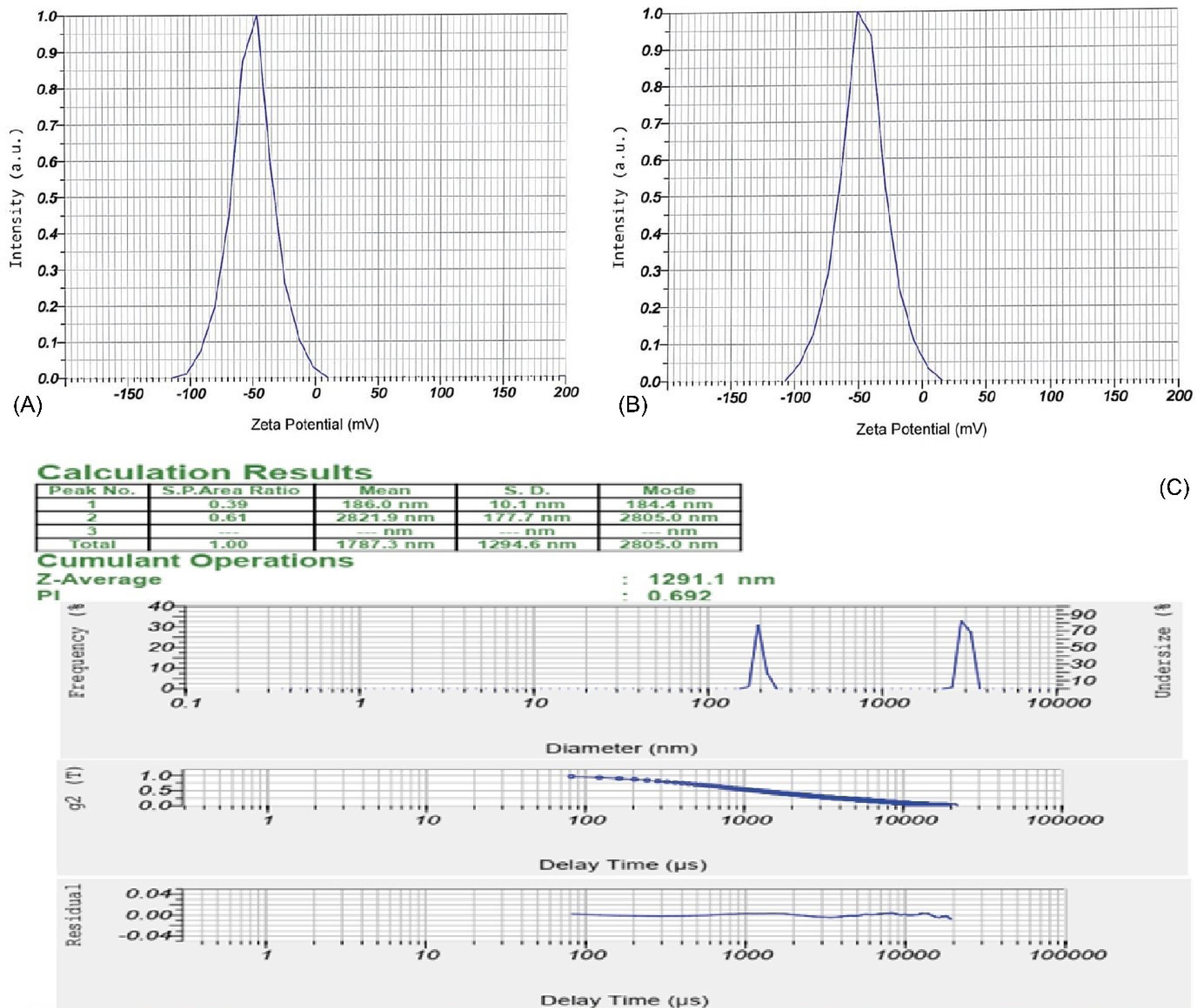
Figure 3.
Zeta Potential of Silver Nanoparticles at Temperatures of 25.1 °C (A) and 25.2 °C (B). These figures display the zeta potential of the synthesized silver nanoparticles at various temperatures, indicating stability and resistance to agglomeration across both values. The data provide a concise description of the zeta potential of the synthesized nanoparticles, showing minimal agglomeration and suitability for various applications. DLS diagram indicates the abundance, hardness, and size of nanoparticles, where the maximum size of the particles is 70-80 nm (C).
.
Zeta Potential of Silver Nanoparticles at Temperatures of 25.1 °C (A) and 25.2 °C (B). These figures display the zeta potential of the synthesized silver nanoparticles at various temperatures, indicating stability and resistance to agglomeration across both values. The data provide a concise description of the zeta potential of the synthesized nanoparticles, showing minimal agglomeration and suitability for various applications. DLS diagram indicates the abundance, hardness, and size of nanoparticles, where the maximum size of the particles is 70-80 nm (C).
Conclusion
The findings of this study indicate that silver nanoparticles synthesized from U. dioica extract possess significant antimicrobial and wound healing properties against S. aureus, E. coli, and Pseudomonas aeruginosa, outperforming the acetonic extract of U. dioica alone. Therefore, green-synthesized silver nanoparticles represent a promising approach for treating burn infections.
Acknowledgments
We would like to express our gratitude to all those who contributed to this study. We would also like to express special thanks to the laboratory technicians and research team members for their invaluable assistance and support in conducting the experiments. Additionally, we extend our appreciation to Azad Zanjan University for providing the facilities necessary for this research.
Competing Interests
The authors declare that they have no conflict of interests.
Funding
No financial support was received for this study.
References
- Girard D, Laverdet B, Buhé V, Trouillas M, Ghazi K, Alexaline MM. Biotechnological management of skin burn injuries: challenges and perspectives in wound healing and sensory recovery. Tissue Eng Part B Rev 2017; 23(1):59-82. doi: 10.1089/ten.TEB.2016.0195 [Crossref] [ Google Scholar]
- Mofazzal Jahromi MA, Sahandi Zangabad P, Moosavi Basri SM, Sahandi Zangabad K, Ghamarypour A, Aref AR. Nanomedicine and advanced technologies for burns: preventing infection and facilitating wound healing. Adv Drug Deliv Rev 2018; 123:33-64. doi: 10.1016/j.addr.2017.08.001 [Crossref] [ Google Scholar]
- Cetik Yildiz S, Demir C, Cengiz M, Ayhanci A. Protective properties of kefir on burn wounds of mice that were infected with S aureus, P auroginasa and E coli. Cell Mol Biol (Noisy-le-grand) 2019; 65(7):60-5. doi: 10.14715/cmb/2019.65.7.11 [Crossref] [ Google Scholar]
- Troitzsch A, Loi VV, Methling K, Zühlke D, Lalk M, Riedel K, et al. Carbon source-dependent reprogramming of anaerobic metabolism in Staphylococcus aureus. J Bacteriol 2021;203(8). doi: 10.1128/jb.00639-20.
- Mahmoudi H, Pourhajibagher M, Alikhani MY, Bahador A. The effect of antimicrobial photodynamic therapy on the expression of biofilm associated genes in Staphylococcus aureus strains isolated from wound infections in burn patients. Photodiagnosis Photodyn Ther 2019; 25:406-13. doi: 10.1016/j.pdpdt.2019.01.028 [Crossref] [ Google Scholar]
- Clegg J, Soldaini E, McLoughlin RM, Rittenhouse S, Bagnoli F, Phogat S. Staphylococcus aureus vaccine research and development: the past, present and future, including novel therapeutic strategies. Front Immunol 2021; 12:705360. doi: 10.3389/fimmu.2021.705360 [Crossref] [ Google Scholar]
- McKay R, Hauk P, Wu HC, Pottash AE, Shang W, Terrell J. Controlling localization of Escherichia coli populations using a two-part synthetic motility circuit: an accelerator and brake. Biotechnol Bioeng 2017; 114(12):2883-95. doi: 10.1002/bit.26391 [Crossref] [ Google Scholar]
- Allocati N, Masulli M, Alexeyev MF, Di Ilio C. Escherichia coli in Europe: an overview. Int J Environ Res Public Health 2013; 10(12):6235-54. doi: 10.3390/ijerph10126235 [Crossref] [ Google Scholar]
- Kunz Coyne AJ, El Ghali A, Holger D, Rebold N, Rybak MJ. Therapeutic strategies for emerging multidrug-resistant Pseudomonas aeruginosa. Infect Dis Ther 2022; 11(2):661-82. doi: 10.1007/s40121-022-00591-2 [Crossref] [ Google Scholar]
- Norbury W, Herndon DN, Tanksley J, Jeschke MG, Finnerty CC. Infection in burns. Surg Infect (Larchmt) 2016; 17(2):250-5. doi: 10.1089/sur.2013.134 [Crossref] [ Google Scholar]
- Church D, Elsayed S, Reid O, Winston B, Lindsay R. Burn wound infections. Clin Microbiol Rev 2006; 19(2):403-34. doi: 10.1128/cmr.19.2.403-434.2006 [Crossref] [ Google Scholar]
- Khalil MA, El Maghraby GM, Sonbol FI, Allam NG, Ateya PS, Ali SS. Enhanced efficacy of some antibiotics in presence of silver nanoparticles against multidrug resistant Pseudomonas aeruginosa recovered from burn wound infections. Front Microbiol 2021; 12:648560. doi: 10.3389/fmicb.2021.648560 [Crossref] [ Google Scholar]
- Mustapha T, Misni N, Ithnin NR, Daskum AM, Unyah NZ. A review on plants and microorganisms mediated synthesis of silver nanoparticles, role of plants metabolites and applications. Int J Environ Res Public Health 2022; 19(2):674. doi: 10.3390/ijerph19020674 [Crossref] [ Google Scholar]
- Kailasa SK, Park TJ, Rohit JV, Koduru JR. Antimicrobial activity of silver nanoparticles. In: Grumezescu AM, ed. Nanoparticles in Pharmacotherapy. William Andrew Publishing; 2019. p. 461-84. doi: 10.1016/b978-0-12-816504-1.00009-0.
- De Vico G, Guida V, Carella F. Urtica dioica (stinging nettle): a neglected plant with emerging growth promoter/immunostimulant properties for farmed fish. Front Physiol 2018; 9:285. doi: 10.3389/fphys.2018.00285 [Crossref] [ Google Scholar]
- Sidor A, Gramza-Michałowska A. Advanced research on the antioxidant and health benefit of elderberry (Sambucus nigra) in food–a review. J Funct Foods 2015; 18(Pt B):941-58. doi: 10.1016/j.jff.2014.07.012 [Crossref] [ Google Scholar]
- Stephen A, Seethalakshmi S. Phytochemical synthesis and preliminary characterization of silver nanoparticles using hesperidin. J Nanosci 2013; 2013(1):126564. doi: 10.1155/2013/126564 [Crossref] [ Google Scholar]
- Akhondzadeh S, Kashani L, Fotouhi A, Jarvandi S, Mobaseri M, Moin M. Comparison of Lavandula angustifolia Mill tincture and imipramine in the treatment of mild to moderate depression: a double-blind, randomized trial. Prog Neuropsychopharmacol Biol Psychiatry 2003; 27(1):123-7. doi: 10.1016/s0278-5846(02)00342-1 [Crossref] [ Google Scholar]
- Abdulsahib SS. Synthesis, characterization and biomedical applications of silver nanoparticles. Biomedicine 2021; 41(2):458-64. doi: 10.51248/.v41i2.1058 [Crossref] [ Google Scholar]
- Kowalska-Krochmal B, Dudek-Wicher R. The minimum inhibitory concentration of antibiotics: methods, interpretation, clinical relevance. Pathogens 2021; 10(2):165. doi: 10.3390/pathogens10020165 [Crossref] [ Google Scholar]
- Dai T, Tegos GP, Lu Z, Huang L, Zhiyentayev T, Franklin MJ. Photodynamic therapy for Acinetobacter baumannii burn infections in mice. Antimicrob Agents Chemother 2009; 53(9):3929-34. doi: 10.1128/aac.00027-09 [Crossref] [ Google Scholar]
- Nasrizal AA, Lowell JJ, Idris J, Nazaruddin AT, Bolong N. The application of statistical ANOVA, LSD and RSM to Agro-based filter design optimization. In: Othman IK, Mohd Haniffah MR, Jamal MH, eds. Proceedings of the 5th International Conference on Water Resources (ICWR). Singapore: Springer; 2023. p. 199-209. doi: 10.1007/978-981-99-3577-2_14.
- Posluszny JA Jr, Conrad P, Halerz M, Shankar R, Gamelli RL. Surgical burn wound infections and their clinical implications. J Burn Care Res 2011; 32(2):324-33. doi: 10.1097/BCR.0b013e31820aaffe [Crossref] [ Google Scholar]
- D’Abbondanza JA, Shahrokhi S. Burn infection and burn sepsis. Surg Infect (Larchmt) 2021; 22(1):58-64. doi: 10.1089/sur.2020.102 [Crossref] [ Google Scholar]
- Ahmad S, Munir S, Zeb N, Ullah A, Khan B, Ali J. Green nanotechnology: a review on green synthesis of silver nanoparticles - an ecofriendly approach. Int J Nanomedicine 2019; 14:5087-107. doi: 10.2147/ijn.S200254 [Crossref] [ Google Scholar]
- Wasef LG, Shaheen HM, El-Sayed YS, Shalaby TI, Samak DH, Abd El-Hack ME. Effects of silver nanoparticles on burn wound healing in a mouse model. Biol Trace Elem Res 2020; 193(2):456-65. doi: 10.1007/s12011-019-01729-z [Crossref] [ Google Scholar]
- Bamal D, Singh A, Chaudhary G, Kumar M, Singh M, Rani N. Silver nanoparticles biosynthesis, characterization, antimicrobial activities, applications, cytotoxicity and safety issues: an updated review. Nanomaterials (Basel) 2021; 11(8):2086. doi: 10.3390/nano11082086 [Crossref] [ Google Scholar]
- Jagtap RR, Garud A, Warude B, Puranik SS. Embelin isolated from Embeliaribes derived silver nanoparticles and its application in breast cancer nanomedicine. Mater Today Proc 2023; 73(Pt 3):403-11. doi: 10.1016/j.matpr.2022.09.265 [Crossref] [ Google Scholar]
- Hassan Afandy H, Sabir DK, Aziz SB. Antibacterial activity of the green synthesized plasmonic silver nanoparticles with crystalline structure against gram-positive and gram-negative bacteria. Nanomaterials (Basel) 2023; 13(8):1327. doi: 10.3390/nano13081327 [Crossref] [ Google Scholar]
- Ansari M, Ahmed S, Abbasi A, Khan MT, Subhan M, Bukhari NA. Plant mediated fabrication of silver nanoparticles, process optimization, and impact on tomato plant. Sci Rep 2023; 13(1):18048. doi: 10.1038/s41598-023-45038-x [Crossref] [ Google Scholar]
- Tesfaye M, Gonfa Y, Tadesse G, Temesgen T, Periyasamy S. Green synthesis of silver nanoparticles using Vernonia amygdalina plant extract and its antimicrobial activities. Heliyon 2023; 9(6):e17356. doi: 10.1016/j.heliyon.2023.e17356 [Crossref] [ Google Scholar]
- Kamble S, Agrawal S, Cherumukkil S, Sharma V, Jasra RV, Munshi P. Revisiting zeta potential, the key feature of interfacial phenomena, with applications and recent advancements. ChemistrySelect 2022; 7(1):e202103084. doi: 10.1002/slct.202103084 [Crossref] [ Google Scholar]
- Alzubaidi AK, Al-Kaabi WJ, Ali AA, Albukhaty S, Al-Karagoly H, Sulaiman GM. Green synthesis and characterization of silver nanoparticles using flaxseed extract and evaluation of their antibacterial and antioxidant activities. Appl Sci 2023; 13(4):2182. doi: 10.3390/app13042182 [Crossref] [ Google Scholar]
- Netala VR, Hou T, Sana SS, Li H, Zhang Z. Rosmarinic acid-rich Perilla frutescens extract-derived silver nanoparticles: a green synthesis approach for multifunctional biomedical applications including antibacterial, antioxidant, and anticancer activities. Molecules 2024; 29(6):1250. doi: 10.3390/molecules29061250 [Crossref] [ Google Scholar]
- Casals-Pascual C, González A, Vázquez-Baeza Y, Song SJ, Jiang L, Knight R. Microbial diversity in clinical microbiome studies: sample size and statistical power considerations. Gastroenterology 2020; 158(6):1524-8. doi: 10.1053/j.gastro.2019.11.305 [Crossref] [ Google Scholar]
- Batool M, Galloway-Peña J. Clinical metagenomics-challenges and future prospects. Front Microbiol 2023; 14:1186424. doi: 10.3389/fmicb.2023.1186424 [Crossref] [ Google Scholar]
- Andrews JM. Determination of minimum inhibitory concentrations. J Antimicrob Chemother 2001; 48 Suppl 1:5-16. doi: 10.1093/jac/48.suppl_1.5 [Crossref] [ Google Scholar]
- Wiegand I, Hilpert K, Hancock RE. Agar and broth dilution methods to determine the minimal inhibitory concentration (MIC) of antimicrobial substances. Nat Protoc 2008; 3(2):163-75. doi: 10.1038/nprot.2007.521 [Crossref] [ Google Scholar]
- Dağlıoğlu Y, Öztürk BY, Khatami M. Apoptotic, cytotoxic, antioxidant, and antibacterial activities of biosynthesized silver nanoparticles from nettle leaf. Microsc Res Tech 2023; 86(6):669-85. doi: 10.1002/jemt.24306 [Crossref] [ Google Scholar]
- Zeinali Aghdam S, Minaeian S, Sadeghpour Karimi M, Tabatabaee Bafroee AS. The antibacterial effects of the mixture of silver nanoparticles with the shallot and nettle alcoholic extracts. J Appl Biotechnol Rep 2019; 6(4):158-64. doi: 10.29252/jabr.06.04.05 [Crossref] [ Google Scholar]
- Hashem AH, Salem SS. Green and ecofriendly biosynthesis of selenium nanoparticles using Urtica dioica (stinging nettle) leaf extract: antimicrobial and anticancer activity. Biotechnol J 2022; 17(2):e2100432. doi: 10.1002/biot.202100432 [Crossref] [ Google Scholar]
- Ebrahiminezhad A, Zare-Hoseinabadi A, Berenjian A, Ghasemi Y. Green synthesis and characterization of zero-valent iron nanoparticles using stinging nettle (Urtica dioica) leaf extract. Green Process Synth 2017; 6(5):469-75. doi: 10.1515/gps-2016-0133 [Crossref] [ Google Scholar]