Avicenna Journal of Clinical Microbiology and Infection. 10(4):152-156.
doi: 10.34172/ajcmi.3499
Original Article
Seroprevalence and Risk Factors Associated With Human Cystic Echinococcosis in Baneh, West of Iran
Mohammad Moghimi 1  , Mehran Bakhtiari 1, Sepehr Matini 1, 2, Mohammad Matini 3, *
Author information:
1Students Research Center, Hamadan University of Medical Sciences, Hamadan, Iran
2Lorestan University of Medical Sciences, Khorramabad, Iran
3Department of Medical Parasitology and Mycology, School of Medicine, Hamadan University of Medical Sciences, Hamadan, Iran
Abstract
Background: Cystic echinococcosis (CE) is a zoonotic helminth infection that has great health and economic importance worldwide. Iran is considered one of the endemic areas of this parasitic infection. The aim of this study was to determine the seroprevalence and risk factors of human CE in Baneh, west of Iran.
Methods: From March to May 2022, 460 individuals who attended health centers in Baneh were tested for the anti-Echinococcus immunoglobulin G (IgG) antibody by enzyme-linked immunosorbent assay. The data were analyzed by Chi-square and binary logistic regression tests.
Results: Fourteen (3.04%) participants (95% CI: 1.49–4.59%) had the anti-Echinococcus IgG antibody. The average (±standard deviation) age of participants was 40.9 (16.8) years, and most of them belonged to the age group of 35–49 years (33.9%) with the highest rate of infection (4.5%). In terms of other demographic variables, seropositivity to CE was higher in women (3.8%) and people who lived in rural areas (5.1%), were housewives (3.6%), and were illiterate (4%) (P>0.05). Binary logistic regression analysis revealed that only keeping dogs at home was a risk factor for CE (adjusted odds ratio: 31.407; 95% CI: 5.343–184.590%; P<0.001).
Conclusion: Direct contact with dogs is the main method of CE transmission. Therefore, public health education and the deworming of dogs can be effective in preventing and controlling CE.
Keywords: Human cystic echinococcosis, Risk factors, Dogs, Echinococcus granulosus, Seroprevalence, Iran
Copyright and License Information
© 2023 The Author(s); Published by Hamadan University of Medical Sciences.
This is an open-access article distributed under the terms of the Creative Commons Attribution License (
https://creativecommons.org/licenses/by/4.0), which permits unrestricted use, distribution, and reproduction in any medium, provided the original work is properly cited.
Please cite this article as follows: Moghimi M, Bakhtiari M, Matini S, Matini M. Seroprevalence and risk factors associated with human cystic echinococcosis in Baneh, West of Iran. Avicenna J Clin Microbiol Infect. 2023; 10(4):152-156. doi:10.34172/ajcmi.3499
Introduction
Cystic echinococcosis (CE) is a zoonotic helminth infection caused by the larval form of Echinococcus granulosussensulato. Humans can be involved as accidental intermediate hosts when eating eggs excreted from a dog as a definitive host (1-4). Echinococcosis has a global distribution and has affected more than 1 million people, with an annual estimation of 19,300 deaths (5). In highly endemic areas, human CE can occur in more than 50 cases per 100 000 people per year, with a prevalence rate of 5%–10% in regions of Peru, Argentina, China, East Africa, and Central Asia (6).
Iran is one of the endemic regions of CE, where 5%–49% of stray dogs and 0.7%–67.7% of slaughtered livestock are infected with E. granulosus(7). In addition, cases of human CE are estimated between 1.18 and 3 per 100 000 populations (8). CE is a serious public health and economic concern in Iran, but the different aspects of its socio-economic effects are not clearly known (9,10). The annual cost of CE is estimated at US$ 232.3 million, including the treatment costs of patients and livestock production losses. The cost of surgical care for liver and lung CE is estimated to be US$ 1027 and US$ 851 per case, respectively. The total cost of human CE is also expected to reach US$ 93.39 million annually. This is mostly due to the effect of reducing human productivity in the asymptomatic and untreated population (8). Several seroepidemiological studies have been performed in the endemic areas to assess the prevalence of CE. Based on a systematic review in 2019, the pooled prevalence of CE in Iran was about 5% (11).
Baneh in Kurdistan province is a pastoral region and one of the endemic areas of CE, with a prevalence rate of up to 5.69% in slaughtered livestock (12). Nonetheless, there is no information about the prevalence of human CE and the main ways of transmission of the infection to humans in this region. Filling information gaps about various aspects of the disease, including complementing human CE data and potential risk factors, is essential to overcome the challenges facing CE control programs in Iran (13). To address this issue, the present study was performed to investigate the seroprevalence and risk factors associated with human CE among people referring to Baneh health centers.
Materials and Methods
Study Area and Sample Collection
This cross-sectional study was conducted in Baneh, a bordering town in western Iran (Figure 1). It is located in the northwest of Kurdistan province at longitude and latitude of 45° 53’ E and 35° 59’ N, respectively. Its altitude is 1,525 meters above sea level. According to the 2016 National Population and Housing Census, the total population of Baneh was 110 218 people.
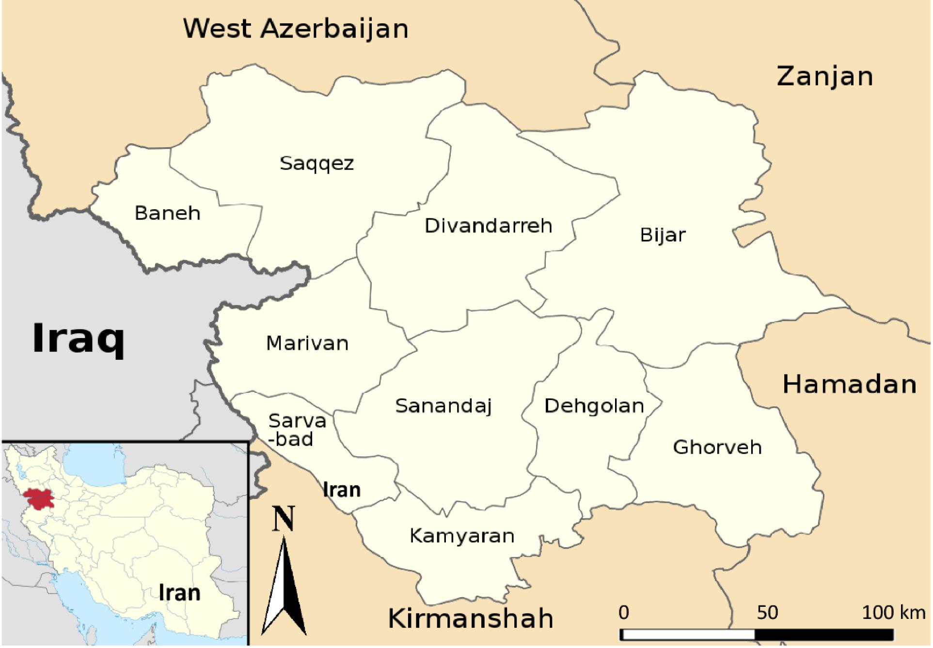
Figure 1.
Map of Iran Showing Kurdistan Province and its Counties
.
Map of Iran Showing Kurdistan Province and its Counties
Ethical Issues and Participants’ Consent
The methodology of this research was approved by the Research Ethics Committee of the Hamadan University of Medical Sciences with approval number IR.UMSHA.REC.1399.850. Written informed consent forms were obtained from the participants attending three health centers in Baneh, including Salahuddin Ayubi hospital, Razi hospital, and the Central Laboratory of Baneh County Health Center. Then, the socio-demographic characteristics of participants and information related to the epidemiology of CE were recorded in separate checklists for each individual. Overall, 460 blood samples were randomly collected from the individuals from March to May 2022. Sera were separated and stored at -70 °C until antibody analysis by the enzyme-linked immunosorbent assay (ELISA).
Enzyme-linked immunosorbent Assay Test
The serum samples were investigated for the anti-Echinococcus immunoglobulin G (IgG) antibody using the Echinococcus IgG ELISA Kit (Pishtaz Teb, Iran). The ELISA was performed in accordance with the manufacturer’s protocol. The color intensity of wells was determined by an ELISA reader at 450 nm. The results were interpreted, and samples with values 10% higher than the cut-off value were considered positive for the specific anti-Echinococcus antibody. Samples with values less than 10% of the cut-off value were considered negative for the antibody. Values between these two values were considered suspicious and re-tested after 2–4 weeks with a fresh specimen.
Data Analysis
The data were analyzed using SPSS software (version 16). The Chi-square test was used to find the association between dependent and independent variables. Statistically significant variables were analyzed by binary logistic regression to predict the risk factors associated with CE. A P value less than 0.05 was considered statistically significant.
Results
Anti-Echinococcus IgG antibody was detected in 14 out of the 460 sera, corresponding to a seroprevalence rate of 3.04% (95% CI:1.49–4.59%). The age range of the participants was from 2 to 99 years, with a mean (standard deviation) age of 40.9 (16.8) years. About 33.9% of the participants belonged to the age group of 35–49 years with the highest rate of infection (4.5%). The related data are provided in Table 1. The number of females was higher than that of males, and their seropositivity to CE (3.8%) was higher than that of males (1.4%). In terms of education, more seropositive cases were found among illiterate subjects (4.0%). The subjects who lived in rural areas and were homemakers had the highest seroprevalence rate in their groups, with 5.1% and 3.6%, respectively. Eight (50.0%) and seven (33.3%) individuals who kept dogs at their homes and had contact with dogs, respectively, were diagnosed as seropositive. In terms of washing vegetables, most of the participants used only water (33.9%), but the highest seropositivity rate (6.9%) was observed in the group that used disinfectant solution. The relationship between seropositivity to CE and contact with dogs and keeping dogs at home was statistically significant (P< 0.001). No statistical correlation was observed between CE seropositivity and other variables (P> 0.05). The adjusted and unadjusted odds ratios of the significant variables are presented in Table 2. Binary logistic regression analysis indicated that keeping dogs at home is an important predictor of CE seropositivity.
Table 1.
Seroprevalence of Cystic Echinococcosis According to Epidemiological Factors in Baneh, Iran
|
Variables
|
Results of Anti-Echinococcus Antibody Test
|
|
P
Value
|
|
Positive
|
Negative
|
Total
|
| Age (y) |
|
|
|
|
| < 20 |
0 (0) |
37 (100) |
37 (8.0) |
0.204 |
| ≥ 20, < 35 |
1 (0.8) |
128 (99.2) |
129 (28.0) |
| ≥ 35, < 50 |
7 (4.5) |
149 (95.5) |
156 (33.9) |
| ≥ 50, < 65 |
4 (4.3) |
89 (95.7) |
93 (20.2) |
| ≥ 65 |
2 (4.4) |
43 (95.6) |
45 (9.8) |
| Gender |
|
|
|
|
| Male |
2 (1.4) |
141 (98.6) |
143 (31.1) |
0.243 |
| Female |
12 (3.8) |
305 (96.2) |
317 (68.9) |
| Education |
|
|
|
|
| Illiterate |
6 (4.0) |
145 (96.0) |
151 (32.8) |
0.672 |
| Primary school |
2 (3.9) |
49 (96.1) |
51 (11.1) |
| Secondary school |
6 (2.5) |
231 (97.5) |
237 (51.5) |
| College |
0 (0) |
21 (100) |
21 (4.6) |
| Occupation |
|
|
|
|
| Manual worker |
5 (3.4) |
141 (96.6) |
146 (31.7) |
0.678 |
| Homemaker |
9 (3.6) |
241 (96.4) |
250 (54.3) |
| Student |
0 (0) |
52 (100) |
52 (11.3) |
| Employee |
0 (0) |
12 (100) |
12 (2.6) |
| Residence |
|
|
|
|
| Urban |
9 (2.5) |
352 (97.5) |
361 (78.5) |
0.193 |
| Rural |
5 (5.1) |
94 (94.9) |
99 (21.5) |
| Contact with dogs |
|
|
|
|
| Yes |
7 (33.3) |
14 (66.7) |
21 (4.6) |
≤ 0.001 |
| No |
7 (1.6) |
432 (98.4) |
439 (95.4) |
| Keeping dogs at home |
| Yes |
8 (50.0) |
8 (50.0) |
16 (3.5) |
≤ 0.001 |
| No |
6 (1.4) |
438 (98.6) |
444 (96.5) |
| Method of washing vegetables |
| Water |
5 (3.2) |
151 (96.8) |
156 (33.9) |
0.185 |
| Salt water solution |
3 (2.2) |
136 (97.8) |
139 (30.2) |
| Detergent solution |
1 (1.1) |
92 (98.9) |
93 (20.2) |
| Disinfectant solution |
5 (6.9) |
67 (93.1) |
72 (15.7) |
Note: Data are expressed as number (percent).
Table 2.
Binary Logistic Regression for Unadjusted (Crude) and Adjusted Odds Ratio for Risk Factors Associated With Cystic Echinococcosis in Baneh
|
Variables
|
Sero- positive
|
Sero- negative
|
Total
|
Model A (cOR)
|
Model B (aOR)
|
|
OR
|
P
Value
|
95% CI
|
OR
|
P
Value
|
95% CI
|
| Keeping dogs at home |
8 |
8 |
16 |
73 |
< 0.001 |
20.52-259.58 |
31.407 |
< 0.001 |
5.343-184.59 |
| Contact with dogs |
7 |
14 |
21 |
30.85 |
< 0.001 |
9.52-99.92 |
3.421 |
0.19 |
0.54-21.55 |
Note. aOR: Adjusted odds ratio; cOR: Crude odds ratio; CI: Confidence interval.
Discussion
CE is considered one of the 17 neglected tropical diseases reported by the World Health Organization, and this helminth zoonotic disease is an important public health problem occurring worldwide (14). Kurdistan province is one of the important areas of animal husbandry in Iran. Therefore, there is a great potential for establishing the life cycle of the parasite in animal hosts and human infections in this region. Despite extensive studies on CE in the country, there is little information about Kurdistan province. The lack of information about the epidemiology of CE in this region justifies the present study conducted in Baneh.
Based on a systematic review performed in 2016, the pooled seroprevalence of CE was estimated to be 6%, with data ranging from 0.23% in Tehran to 15.4% in Khorramabad (15). In the present study, 3.04% of the participants were seropositive for the anti-Echinococcus antibody. This value is within the range of those reported in the systematic review study. Nonetheless, the seroprevalence of CE in Baneh was lower than in regions such as Khuzestan (13.8%) (16), Yasuj (7.2%) (17) in the south of the country, Lorestan (15.4%) (18) in the center of Iran, but more than in regions such as Qom (1.6%) (19), Arak (1.3%) (20), Isfahan (1.1%) (21) in central semi-arid areas, and East Azerbaijan (1.28%) (22) in the northwest of Iran. Further, the prevalence rate was higher than the result of the study by Nazari et al (2.2%). This study was conducted in 2019 in Sanandaj, the capital of Kurdistan province (23). The seroprevalence of CE can be affected by some factors such as test method, sample size, geographical area, population under study, and demographic factors, partly explaining the difference in various studies.
Immunoassay based on ELISA techniques is a reliable tool for epidemiological surveys of infectious diseases. ELISA with the AgB of E. granulosus has high sensitivity and specificity for the detection of CE (10). Therefore, in this study, AgB-ELISA was used for the detection of IgG antibodies against Echinococcus.
In the current study, the highest CE seropositivity was detected in the 35–49 age group, and all individuals < 20 years were seronegative. Although this finding was not significant, it is in line with the results of other studies conducted in Iran (10,15). In the study performed in Fars province, the relationship between seropositivity and age was significant, and multivariate logistic regression analysis showed that the age group of 35–45 years was associated with seropositivity to CE (OR: 2.23, P= 0.002) (10). Despite the majority of studies, Sarkari et al reported a prevalence of 7.6% among children aged 5–20 years in rural Fars province (24). Naturally, age increases the likelihood of exposure to the parasite, and the prevalence of the infection is expected to increase with age (25).
The results of our study demonstrated that the prevalence rate in women (3.8%) is higher than in men (1.4%) (P> 0.05). The findings of some studies are consistent with those of our study (10,16,25,26), while other studies have reported conflicting results (19-21). Additionally, lifestyle can affect the prevalence rate of infection. The traditional lifestyle is still observed in most parts of Iran, including the Kurdistan region. In this lifestyle, housewives are involved in activities such as cleaning the home and neighborhood environment, washing vegetables, caring for animals, and growing vegetables in the yard, allowing them to have more exposure to possible sources of infection.
In the present study, 43.9% of the participants were illiterate or had primary education, which had the highest CE prevalence of 4.0% and 3.9%, respectively. This finding is also in line with that of another study (20). Other findings of our study revealed that seropositive individuals in this region had poor knowledge and attitudes toward this parasitic disease. Obviously, low education and a lack of health awareness are associated with the prevalence of infectious diseases (27,28).
In Baneh, some people reside in the village for some time of the year due to agricultural or livestock farming activities. Furthermore, 21.5% of the participants were only residents of the village, and the prevalence rate among them (5.1%) was more than twice that of the residents in the city (2.5%) (P > 0.05). In our study, keeping dogs at home and contact with dogs were significantly associated with seropositivity to CE. Moreover, the binary logistic regression test represented that people who kept dogs at home had a 31.4 times higher chance of seropositivity to CE compared to people who did not keep dogs. Studies conducted in Iran indicated a significant prevalence of echinococcosis in dogs (7,29,30). Dogs, as definitive hosts, are the most important sources of infection for humans because they spread the parasite’s eggs through their feces in the environment. Swallowing the eggs through food, water, or contact with dogs are the most important ways of human infection (1,27,28). Rural lifestyles have often been suggested as a set of risk factors for human CE. Employment in agriculture and animal husbandry, contact with dogs, and a low level of hygiene facilitate the conditions for the spread of human CE in villages (27,28,30).
Finally, there are limitations to seroepidemiological studies that should be taken into consideration. The diagnosis of CE is mainly based on a combination of an imaging approach and serological methods as auxiliary tests. Limitations in the sensitivity and specificity of serological methods due to the location of the cyst lesion and cross-reactions with other helminth infections or non-infectious conditions can lead to underestimating or overestimating the prevalence of CE (1).
Conclusion
The considerable seroprevalence of CE among the individuals indicated that CE is a major health problem in Baneh. Contact with dogs is the main route of echinococcosis transmission in this region, and people lack the necessary information about this parasitic disease. Therefore, the prevention and control of CE transmission are considered priorities in this region. In addition to taking health measures to control echinococcosis, creating educational campaigns to raise people’s awareness of this infection and deworming dogs can play an effective role in controlling human CE.
Acknowledgements
The authors are grateful to all participants for their cooperation in this research. In addition, we would like to thank the Vice-chancellor of Research and Technology, Hamadan University of Medical Sciences, for their financial support (Project No. 9910237367).
Authors’ Contribution
Conceptualization: Mehran Bakhtiari, Mohammad Matini.
Data curation: Mohammad Matini, Mehran Bakhtiari.
Formal analysis: Mohammad Matini, Sepehr Matini.
Funding acquisition: Mohammad Matini.
Investigation: Mohammad Moghimi, Sepehr Matini, Mehran Bakhtiari.
Methodology: Mohammad Matini, Mohammad Moghimi.
Project administration: Mohammad Matini.
Resources: Mohammad Matini.
Supervision: Mohammad Matini.
Visualization: Mohammad Matini.
Writing–original draft: Sepehr Matini.
Writing–review & Editing: Mohammad Matini.
Competing Interests
The authors declare no conflict of interests.
Ethical Approval
This study was approved by the Research Ethics Committee of the Hamadan University of Medical Sciences (Ethical code: IR.UMSHA.REC.1399.850).
Funding
This study was funded by the Vice-chancellor of Research and Technology, Hamadan University of Medical Sciences(Project No. 9910237367).
References
- Agudelo Higuita NI, Brunetti E, McCloskey C. Cystic echinococcosis. J Clin Microbiol 2016; 54(3):518-23. doi: 10.1128/jcm.02420-15 [Crossref] [ Google Scholar]
- Moro P, Schantz PM. Echinococcosis: a review. Int J Infect Dis 2009; 13(2):125-33. doi: 10.1016/j.ijid.2008.03.037 [Crossref] [ Google Scholar]
- Manzano-Román R, Sánchez-Ovejero C, Hernández-González A, Casulli A, Siles-Lucas M. Serological diagnosis and follow-up of human cystic echinococcosis: a new hope for the future?. Biomed Res Int 2015; 2015:428205. doi: 10.1155/2015/428205 [Crossref] [ Google Scholar]
- Matini M, Fallah M, Maghsood AH, Saidijam M, Fasihi Harandi M. Echinococcus granulosus sensu stricto in livestock and human in Hamadan, western Iran. Iran J Parasitol 2019; 14(2):288-96. [ Google Scholar]
- World Health Organization (WHO). WHO Estimates of the Global Burden of Foodborne Diseases: Foodborne Disease Burden Epidemiology Reference Group 2007-2015. Geneva: WHO; 2015. Available from: https://apps.who.int/iris/handle/10665/199350.
- World Health Organization (WHO). Echinococcosis Fact Sheet. Geneva: WHO; 2020. Available from: https://www.who.int/news-room/fact-sheets/detail/echinococcosis. Accessed January 5, 2021.
- Rokni MB. The present status of human helminthic diseases in Iran. Ann Trop Med Parasitol 2008; 102(4):283-95. doi: 10.1179/136485908x300805 [Crossref] [ Google Scholar]
- Fasihi Harandi M, Budke CM, Rostami S. The monetary burden of cystic echinococcosis in Iran. PLoS Negl Trop Dis 2012; 6(11):e1915. doi: 10.1371/journal.pntd.0001915 [Crossref] [ Google Scholar]
- Matini M, Roostaei M, Fallah M, Maghsood AH, Saidijam M, Fasihi Harandi M. Genetic identification of Echinococcus granulosus isolates in Hamadan, western Iran. Iran J Parasitol 2018; 13(3):423-9. [ Google Scholar]
- Safarpour AR, Omidian M, Pouryousef A, Fattahi MR, Sarkari B. Serosurvey of cystic echinococcosis and related risk factors for infection in Fars province, southern Iran: a population-based study. Biomed Res Int 2022; 2022:3709694. doi: 10.1155/2022/3709694 [Crossref] [ Google Scholar]
- Mahmoudi S, Mamishi S, Banar M, Pourakbari B, Keshavarz H. Epidemiology of echinococcosis in Iran: a systematic review and meta-analysis. BMC Infect Dis 2019; 19(1):929. doi: 10.1186/s12879-019-4458-5 [Crossref] [ Google Scholar]
- Yakhchali M, Ghargi B. A survey on prevalence of hydatidosis in slaughtered ruminant in Baneh (Kurdistan province) in 2001. Iran Vet J 2006; 10(12):88-95. [ Google Scholar]
- Ebrahimipour M, Budke CM, Fasihi Harandi M. Control of cystic echinococcosis in Iran: where do we stand?. Trends Parasitol 2020; 36(7):578-81. doi: 10.1016/j.pt.2020.04.007 [Crossref] [ Google Scholar]
- Tao J, Du X, Liu K, Wang C, Lv Y, Wang M. Clinical characteristics and antibodies against Echinococcus granulosus recombinant antigen P29 in patients with cystic echinococcosis in China. BMC Infect Dis 2022; 22(1):609. doi: 10.1186/s12879-022-07597-8 [Crossref] [ Google Scholar]
- Shafiei R, Hosseini Teshnizi S, Kalantar K, Gholami M, Mirzaee G, Mirzaee F. The seroprevalence of human cystic echinococcosis in Iran: a systematic review and meta-analysis study. J Parasitol Res 2016; 2016:1425147. doi: 10.1155/2016/1425147 [Crossref] [ Google Scholar]
- Rafiei A, Hemadi A, Maraghi S, Kaikhaei B, Craig PS. Human cystic echinococcosis in nomads of south-west Islamic Republic of Iran. East Mediterr Health J 2007; 13(1):41-8. [ Google Scholar]
- Sarkari B, Sadjjadi SM, Beheshtian MM, Aghaee M, Sedaghat F. Human cystic echinococcosis in Yasuj district in southwest of Iran: an epidemiological study of seroprevalence and surgical cases over a ten-year period. Zoonoses Public Health 2010; 57(2):146-50. doi: 10.1111/j.1863-2378.2008.01200.x [Crossref] [ Google Scholar]
- Zibaei M, Azargoon A, Ataie-Khorasgani M, Ghanadi K, Sadjjadi SM. The serological study of cystic echinococcosis and assessment of surgical cases during 5 years (2007-2011) in Khorram Abad, Iran. Niger J Clin Pract 2013; 16(2):221-5. doi: 10.4103/1119-3077.110156 [Crossref] [ Google Scholar]
- Rakhshanpour A, Fasihi Harandi M, Moazezi S, Rahimi M, Mohebali M, Mowlavi G. Seroprevalence of human hydatidosis using ELISA method in Qom province, Central Iran. Iran J Parasitol 2012; 7(3):10-5. [ Google Scholar]
- Darabi F, Bakhtiari M, Matini S, Matini M. Seroepidemiology of hydatid cyst in outpatients attending health centers in Arak city, Iran, 2020. Avicenna J Clin Med 2022; 28(4):238-243. doi: 10.52547/ajcm.28.4.238 [Crossref] [ Google Scholar]
- Ilbeigi P, Mohebali M, Beigom Kia E, Saber-Inasab M, Aryaeipour M, Bizhani N. Seroepidemiology of human hydatidosis using AgB-ELISA test in Isfahan city and suburb areas, Isfahan province, Central Iran. Iran J Public Health 2015; 44(9):1219-24. [ Google Scholar]
- Garedaghi Y, Bahavarnia SR. Seroepidemiology of human hydatidosis by ELISA method in East-Azarbaijan province in Iran in year 2009. Iran J Epidemiol 2011; 7(2):25-9. [ Google Scholar]
- Nazari N, Nayeri T, Hazrati F. Seroprevalence of human cystic echinococcosis in Sanandaj city, Kurdistan province, Western Iran. Int J Med Lab 2021; 8(3):188-95. doi: 10.18502/ijml.v8i3.7325 [Crossref] [ Google Scholar]
- Sarkari B, Arefkhah N, Ghorbani F, Meskini F, Yektaeian N, Shahriarirad S. Seroprevalence of cystic echinococcosis and related risk factors for infection among children in a rural community in Fars province, southern Iran. Clin Epidemiol Glob Health 2020; 8(1):13-6. doi: 10.1016/j.cegh.2019.03.009 [Crossref] [ Google Scholar]
- Heidari Z, Mohammadi-Ghalehbin B, Alizadeh Z, Molaei S, Peeri Dogaheh H, Mirzanejad-Asl H. Seroprevalence of human hydatidosis in Ardabil province, north-west of Iran. Iran J Parasitol 2021; 16(4):593-600. doi: 10.18502/ijpa.v16i4.7872 [Crossref] [ Google Scholar]
- Wang Q, Huang Y, Huang L, Yu W, He W, Zhong B. Review of risk factors for human echinococcosis prevalence on the Qinghai-Tibet Plateau, China: a prospective for control options. Infect Dis Poverty 2014; 3(1):3. doi: 10.1186/2049-9957-3-3 [Crossref] [ Google Scholar]
- Borhani M, Fathi S, Darabi E, Jalousian F, Simsek S, Ahmed H. Echinococcoses in Iran, Turkey, and Pakistan: old diseases in the new millennium. Clin Microbiol Rev 2021; 34(3):e0029020. doi: 10.1128/cmr.00290-20 [Crossref] [ Google Scholar]
- Mikaeili Galeh T, Spotin A, Mahami-Oskouei M, Carmena D, Rahimi MT, Barac A. The seroprevalence rate and population genetic structure of human cystic echinococcosis in the Middle East: a systematic review and meta-analysis. Int J Surg 2018; 51:39-48. doi: 10.1016/j.ijsu.2018.01.025 [Crossref] [ Google Scholar]
- Rahmati K, Maghsoud AH, Matini M, Motevalli Haghi M, Fallah N, Fallah M. Study of Intestinal helminthes of stray dogs and their public heath importance in Hamadan city. Avicenna J Clin Med 2016; 23(3):214-20. doi: 10.21859/hums-23033 [Crossref] [ Google Scholar]
- Dorjsuren T, Ganzorig S, Dagvasumberel M, Tsend-Ayush A, Ganbold C, Ganbat M. Prevalence and risk factors associated with human cystic echinococcosis in rural areas, Mongolia. PLoS One 2020; 15(7):e0235399. doi: 10.1371/journal.pone.0235399 [Crossref] [ Google Scholar]