Microscopy and its Application in Microbiology and Medicine From Light to Quantum Microscopy: A Mini Review
Avicenna J Clin Microbiol Infect, 6(4), 133-137; DOI:10.34172/ajcmi.2019.24
Review Article
Microscopy and its Application in Microbiology and Medicine From Light to Quantum Microscopy: A Mini Review
Amir Khodavirdipour1,2*, Mahboobeh Mehregan3, Ali Rajabi2, Yasoub Shiri4
1
Division of Human Genetics, Department of Anatomy, St. John’s Hospital, Bangalore, India
2
Department of Biology, Faculty of Natural sciences, University of Tabriz, Tabriz, Tabriz, Iran
3
Department of Biology, Faculty of Science, University of Zabol, Zabol, Iran
4
Assistant Professor of Agronomy and Plant Breeding, Agricultural Research Institute, University of Zabol, Zabol, Iran
*Corresponding author:
Amir Khodavirdipour
Division of Human Genetics,
Department of Anatomy, St.
John’s Hospital, Bangalore,
Karnataka, INDIA
Tel: 0091-9986695070, Email:
amir_kvp@yahoo.com
Abstract
The invention of the microscope was a great help to science to reach a new extension, especially biology. By employing microscopes, the researchers were able to prove new dimensions of living such as a microorganism, work, and cell study, and go in-depth of the minute part of fungi, plants or even the animals. In the mid-20th century, the electron was a great help and opened a new horizon. The scanning microscopes are even more delicate and sophisticated and also have higher magnification power compared to light microscopes. Basically, microscopes utilize the simply visible rays which refract the lenses, X-rays, electron, and infrared rays. In addition, they are designed to detect smaller and smaller particles and structures. Further, scanning electron microscopes are capable of resolving viruses which are extremely smaller than any microorganism and cell, they can enlarge the view of small viruses, which allows the researcher and scientists to develop the cure or a new vaccine for related diseases in animals or even humans. Furthermore, scanning electron microscopes have the power of magnification even till million times, almost molecule size, viruses and also nanoparticles. Moreover, they use software for correcting and enhancing the magnification and resolution of the image. The software helps to view the desired objects even if they are a few molecules thick. This mini-review extensively discusses each type and their different available classifications.
Keywords: Microscope, Compound microscope, Electron microscope, Light microscope, Quantum microscope, X-ray microscope
Background
Let’s begin with the history of the invention of the microscope. Similar to the other inventions, there are disputes about who first invented the microscope. It dates back to the first century when the glass was first invented by the Roman (1). They were working on glass uses and how to observe an object through the glass and enlarge the object by viewing through the glass. Then, Italian Salvino D’Armate was the first person to create eye gear in the 13th century, which provided the individual with magnification to one eye. The earliest very simple form of magnification glass had the power of 6x to 10x and usually used to inspect small insects or a small part of the plants. Later on, two glassmakers from the Netherlands, Zacharias Jansen, and his father started to work and experiment with larger lenses during the 1590s, (1). They lay several lenses in a tube-like structure and made a huge discovery. The sample object near the tube appeared extremely larger than a single lens would do. The power of this device was only around 9x and the images were blurry and not clear. In fact, Jansen’s microscope failed to survive with its low magnification power and blurry image and it was a novelty invention compared to the microscope. Dutch scientist and draper, Anton Van Leeuwenhoek (1632-1723) was one of the main individuals or maybe the first one to make a real microscope in the late 17th century (2). Van Leeuwenhoek nailed greater achievement than his rivals by making a superlens after 550 failed attempts. His last lens was a huge success and had a magnifying power of 270x where his rivals had a maximum of 50x. Moreover, Van Leeuwenhoek made lots of biological discoveries by using this microscope. First, he observed bacteria, yeast, a stream of life in a drop of water, and even blood circulation in capillaries (2). He wrote and sent over a hundred letters to the Royal Society of England and French Academy and reported his findings which included living and non-living samples. His works verified and further continued by English scientist, Robert Hooke, who published the first work on microscope in 1665 by the name Micrographia. All the advancements have so far come to live based on his works. Abbe defined the theoretically minimum size around 200 nm to be viewed by a microscope; since light microscopes are only able to use the wavelength of visible lights to focus on objects and on average, we assume visible light wavelength for 550 nm. In contrast, an electron microscope can magnify the objects thousands of times more than a normal light microscope (3).
Methodology
The present article reviewed studies for current projects based on published works in Medline/PubMed, Scopus and Google scholar for the last 9 years (from January 1, 2010 to December 31, 2019), as well as the references from the above-mentioned papers. The search narrowed in time for meant for the latest and more updated papers. For this latest updated review, our search terms included the “history of the microscope”, “advances in microbiology”, “microscope in medicine and biology”, “optic in biology”, and “microscope application”. Later, all duplicate papers from the above-mentioned databases were discarded and only eligible papers were processed for the next round based on our desired set goals.
Timeline of the History of the Microscope
-
2nd century BC: Claudius Ptolemy defined that a stick can appear bend in a bowl of water.
-
1st century BC: Romans experimenting with glass and accidentally found that objects appeared bigger and larger through some shapes of glasses.
-
12th century AD: Italian Salvino D’Armate made the first magnifying eyepiece (Figure 1).
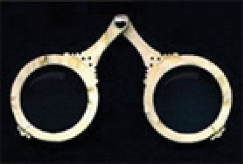
Salvino D’Armates’s Magnifying Eyepiece (1).
Zechariah and His Father, Hans’s First Compound Microscope (1).
Microscope History Timeline
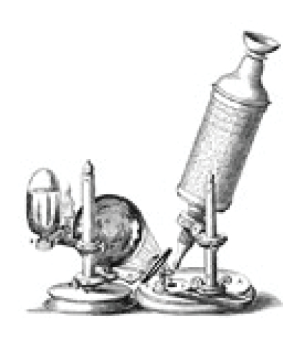
Galilei’s Compound Microscope (2).
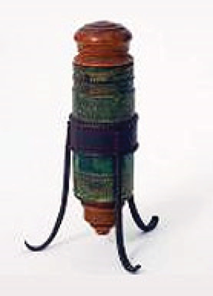
One of Robert Hooke’s Observatory Devices (3).
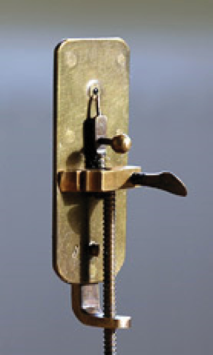
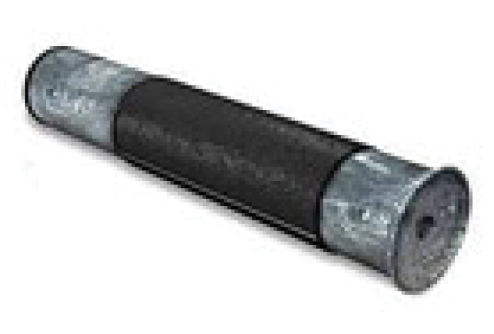
Anton Van Leeuwenhoek’s Lens Grinding Device (2). Figure 6. Joseph Jackson Lister build an achromatic lens that eradicating the chromatic effect made by different light’s wavelengths.
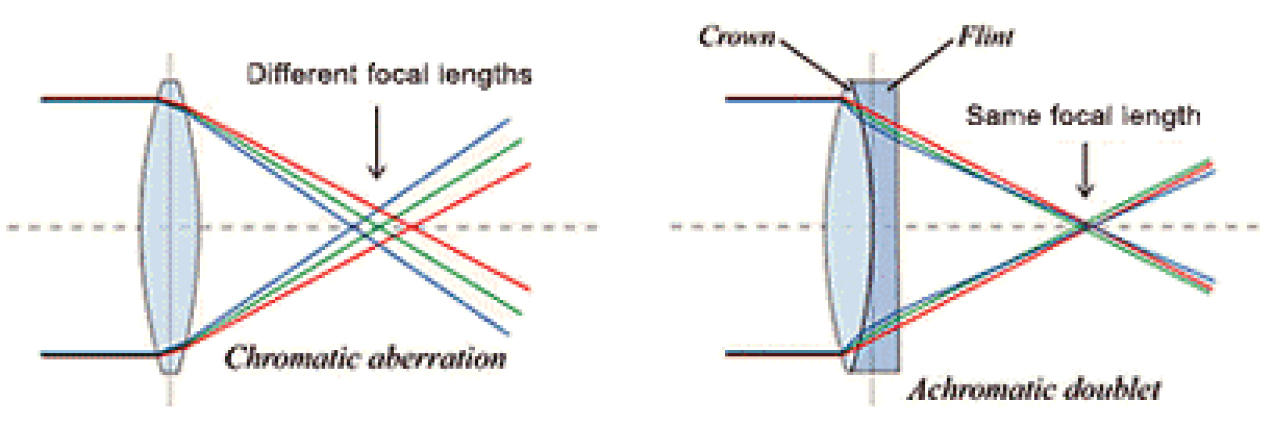
Joseph Jackson Lister build an achromatic lens that eradicating the chromatic
effect made by different light’s wavelengths.
-
The 1860s: Ernst Abbe discovered the Abbe sine condition (a condition that must be achieved by a lens to produce a sharp image), which was a breakthrough in microscope history that was based on trial and error till 1860.
-
1931: Ernst Ruska built the first electron microscope.
Microscopes and its Classifications
Generally, microscopes can be categorized based on some factors like optical function, structure, or application.
Basically, major classification is as follows:
Optical
-
Binocular stereoscopic microscope
-
Bright field microscope
-
Polarizing microscope
-
Phase contrast microscope
-
Differential interference contrast microscope
-
Fluorescence microscope
-
Total internal reflection fluorescence microscope
-
Laser microscope (laser scanning confocal microscope)
-
Multiphoton excitation microscope
-
Structured illumination microscope.
Electron
1. Transmission electron microscope (TEM)
2. Scanning electron microscope (SEM).
Scanning Probe Microscope (SPM)
1. Atomic force microscope (AFM);
2. Scanning near-field optical microscope (SNOM).
Others
1. X-ray microscope;
2. Ultrasonic microscope.
The other categories are based on the structure and application.
Application
1. Biological microscope;
2. Stereoscopic microscope.
Structure
1. Upright microscope;
2. Inverted microscope.
2. Binocular Stereoscopic Microscope
This is a type microscope which allows for easily observing the 3D at not very high magnification.
2.1. Bright-Field Microscope
This microscope utilizes lights which are transmitted to observe at high magnification.
2.2. Polarizing Microscope
The polarizing microscope has different types of lights which transmit the characteristics of materials, including crystalline structures in order to make a perfect image.
2.3. Phase-Contrast Microscope
This device brings to life even minute irregularities by utilizing the interference of the light. It is possible to observe living objects even without staining them.
2.4. Differential Interference Contrast Microscope
This is similar to the above-mentioned microscope but produces higher resolution pictures. However, it has its limitations due to the nature of the lights.
2.5. Fluorescence Microscope
This life science microscope captures fluorescence emitted from the sample by using a dedicated source of light like the mercury lamps. In combination with other equipment, a bright-field microscope also can produce fluorescence images (4).
2.6. Total Internal Reflection Fluorescence Microscope
It is a kind of fluorescent microscope that uses a passing wave only to illuminate the surface of the specimen. The viewed region is generally very thin in comparison to other regular microscopes. In addition, molecular unit observation is possible by using this microscope (4).
2.7. Laser Microscope (Laser Scanning Confocal Microscope)
The laser beam in this microscope is used to clear the picture of samples with changes in focal distances (3).
2.8. Multiphoton Excitation Microscopy
The multiphoton excitation microscope laser causes very little damage to living cells and interestingly allows higher resolution observation of obscure areas. Further, this microscope is usually used to observe the blood flow or nerve cells in the brain (3).
2.9. Structured Illumination Microscopy
This very high-resolution microscope with highly sophisticated technology is utilized to handle the problem of the limited and poor resolution of optical microscopes that are only caused by light diffraction (5).
3. Electron Microscope
These microscopes instead of the light beams emit an electron beam toward an object in order to magnify them.
3.1. Transmission Electron Microscope
A small source at the highest part of the microscope emits the electron which can travel via a vacuum column of the microscope. Instead of the lens which focuses the light in the light microscope, this microscope uses an electromagnetic lens that focuses the electrons into a beam. The electron beam then passes via a specimen. Depending on the density of the material and texture of the object, few electrons disappear from the beam. At the base part of microscope, electrons which remain unscattered collide to the screen, which produces a shadow-like image of the understudy specimen based on its density. The image can be analyzed and photographed for further study by the operator (6).
3.2. Scanning Electron Microscope
High-speed electrons in SEM have outstanding amounts of energy and above energy dispersed in very different signals produced by the electron–specimen interaction at the time the electrons speed decreases in solid objects. This signal may consist of secondary electrons, photons, visible lights, and heat. These electrons and reflexed electrons are commonly used in image production (6).
4. Scanning Probe Microscope
The scanning probe microscope scans the outer layer of an object by a probe and the intended interaction measures the area and identifies its properties (4).
4.1. Atomic Force Microscope
Almost all atomic force microscopes typically use a laser system in which the beam is reflected from behind the reflective atomic force microscope lever and into a detector. Furthermore, atomic force microscopes are mainly fabricated from Si3N4 and their radius is usually from couple to 10 seconds of a nanometer (3).
4.2. Scanning Near-field Optical Microscope (SNOM/ NSOM)
A near-field scanning optical microscope (NSOM) is a microscopy technique to study nanostructure. In SNOM (Scanning near-field optical microscope), the exciting laser beam is focused by a tight aperture less than a wavelength, which results in the far side of the minimum aperture. Then, the specimen is scanned in a tiny distance less than the aperture (5).
5. Others
5.1. X-ray Microscope
An X-ray microscope uses the radiation which is electromagnetic in soft X-ray to make enlarged images of the specimen. As far as X-rays penetrate through almost all subjects, there is no need for any specific staining techniques. Unlike compound or light microscopes, X-ray microscopes do not refract or reflect and they are not visible by our eyes. Nevertheless, an X-ray microscope exhibits a film or uses a charge-coupled device (a detecting device) to identify the X-ray which passes through the object. This is based on contrast and this technology is used by measuring a difference absorption of soft X-ray in the water window region. The wavelength of 2.34-4.4 nm has an energy level of 280 to 530 cV. The main element in the living cell and water uses a carbon atom and oxygen cell, respectively (7).
5.2. Ultrasonic Microscope
Acoustic microscopy is a type of microscope that uses very high or an ultra-high ultrasound frequency. These microscopes can penetrate through almost all solid objects to make them visual to the human eye and make an image of their internal properties, including issues and problems like cracks or the like (7).
Discussion
How These Microscopes Work in General
The optical microscope is the simplest and earliest microscope that uses a single lens, which is a convex lens, to magnify an object under focus. Over the decades, researchers have tried more lenses and created compound microscopes with increased magnification power. Compound microscopes can magnify the objects as small as 0.2 nm visible to the human eye. Furthermore, with the advancement of technologies and the use of different strategies, they become better in any way. Moreover, these microscopes can produce a high degree of magnification but in low resolution and they are more commonly used types of microscopes in basic labs and schools.
Electron Microscope
Electron microscopes emit a beam of electrons at a sample, which is held in a vacuum and airless tube. The researchers typically use these microscopes to analyze and study the cells. In the case of TEM, the emitted electrons via a dehydrated thin sample reach the film behind the sample and produce an image which includes the inner structure of a cell as well. Additionally, the scanning electron microscopes shoot a beam of electrons over the surface of a subject and create a three-dimensional image. These microscopes have a magnification of up to one million times what a human eye can observe with clear resolution.
Scanning Probe Microscope
The scanning probe microscope works based on a probe, whose metallic tip is as the size of an atom. This probe measures many things while it is rolling over the subject. It discovers from magnetic properties and power to physical surface and depth. In addition, these microscopes are outstandingly powerful and can very small objects even less than a nanometer. Nonetheless, the result is in black and white because the probe uses the other sources of the light than the visible light. This type of microscope was invented in 1981 and was first called a scanning tunneling microscope.
Quantum Microscopy
Quantum Microscope as an MRI for Molecules
This type of microscope works based on a diamond, which employs the magnetic resonance of the electrons to identify charged atoms in chemical interactions and reactions in real-time (8). Further, this microscope uses a central sensor which is built out of a 2 mm diamond where allows the scientist to study and analyze topics like how a DNA molecule winds and folds inside a cell and how drugs work inside bacteria or cells. Remarkably, this device can produce images of each individual ion even a liquid solvent and describe the biochemical reaction while happening without intermediating or interrupting the ongoing process (8). The interesting point is that this machine permits the measurement and evaluation of the specimen without touching it, this procedure is also known as the interaction-free process or measurement (9). Scientists long awaited for such an imaging technology in order to observe the molecular structure and interaction that exactly work based on an MRI machine of the hospital, which harmlessly shows the internal structure of the body without any invasive intervention. The current power of quantum MRI is 10 micrometers and above (8). A team at the University of Melbourne, Australia led by Lloyd Hollenberg and David Simpson who were both physicists, invented this device to observe the metallic ions in living cells.
Conclusions
In general, after knowing the pros and cons of all types of microscopes available today, an electron microscope is the most widely used in the field of medicine for precise diagnosis of liver, muscle, intestine, kidney, and nerves. More precisely, these microscopes are further utilized in clinical microbiology, virology, and mycology and specifically become the gold standard in molecular biology.
Ethical Approval
Not applicable.
Conflict of Interest Disclosures
The authors declare that there is no conflict of interests regarding this paper.
References
- Microscope history (blog). http://knowledgein21century.blog. ir/1394/10/20/Microscope-history.
- Egerton FN. 2006. A history of the ecological sciences, part 19: Leeuwenhoek’s microscopic natural history. The Bulletin of the Ecological Society of America 2006;87:47-58. doi:10.1890/0012-9623(2006)87[47:AHOTES]2.0.CO;2. [Crossref]
- An experimental society. HistoryExtra website. https://www. historyextra.com/period/an-experimental-society/. Published January 23, 2010.
- Jang WH, Kwon S, Shim S, Jang WS, Myung JK, Yang S, et al. Comparison between reflectance confocal microscopy and 2-photon microscopy in early detection of cutaneous radiation injury in a mouse model in vivo. J Biophotonics 2018;11(10):e201700337. doi: 10.1002/jbio.201700337. [Crossref]
- Costa EC, Moreira AF, de Melo-Diogo D, Correia IJ. Polyethylene glycol molecular weight influences the cleart2 optical clearing method for spheroids imaging by confocal laser scanning microscopy. J Biomed Opt 2018;23(5):1-11. doi: 10.1117/1. jbo.23.5.055003. [Crossref]
- Allgeier S, Bartschat A, Bohn S, Peschel S, Reichert KM, Sperlich K, et al. 3d confocal laser-scanning microscopy for large-area imaging of the corneal subbasal nerve plexus. Sci Rep 2018;8(1):7468. doi: 10.1038/s41598-018-25915-6. [Crossref]
- Johnson D. What Are the Three Main Types of Microscopes? https://sciencing.com/three-main-types-microscopes-12507. html. Updated April 26, 2018.
- Reardon S. Quantum microscope offers MRI for molecules. Nature News 2017;543:162.
- Baek KI, Ding Y, Chang CC, Chang M, Sevag Packard RR, Hsu JJ, et al. Advanced microscopy to elucidate cardiovascular injury and regeneration: 4D light-sheet imaging. Prog Biophys Mol Biol 2018;138:105-15. doi: 10.1016/j. pbiomolbio.2018.05.003. [Crossref]